Spongy Bone Drawing
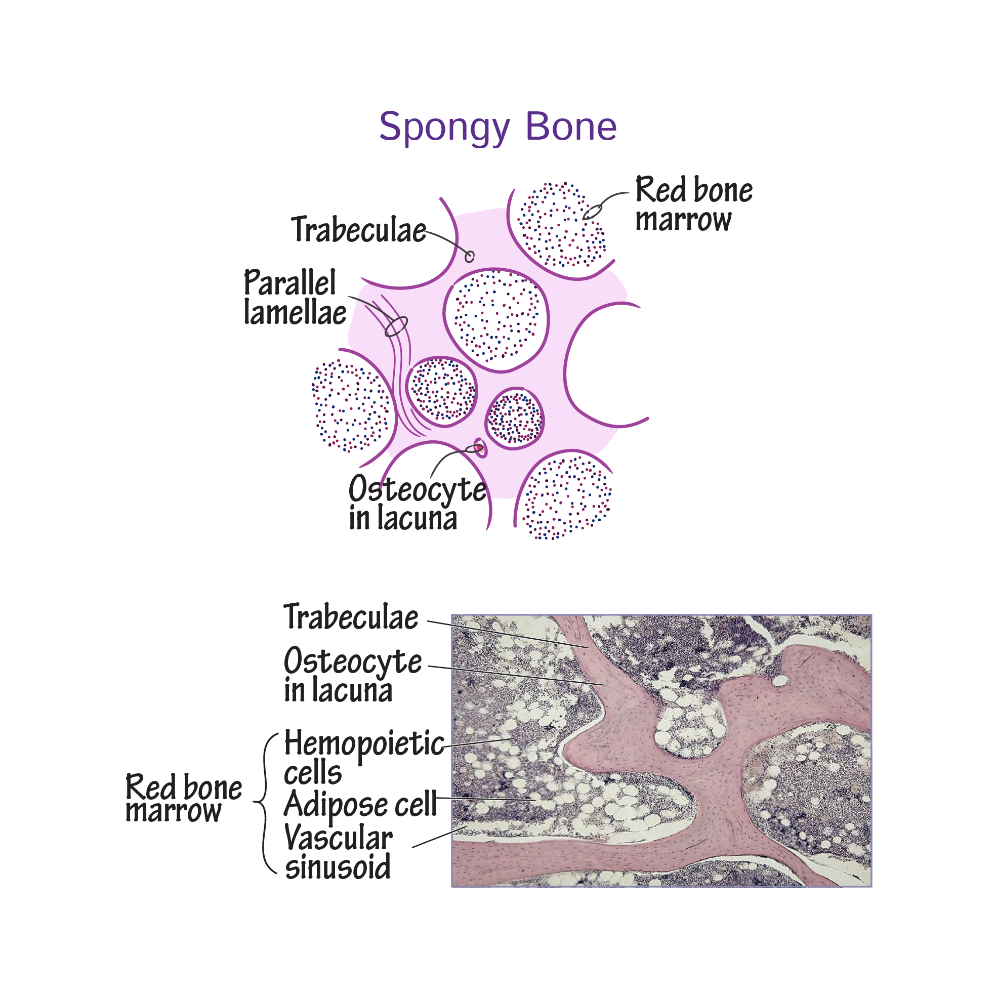
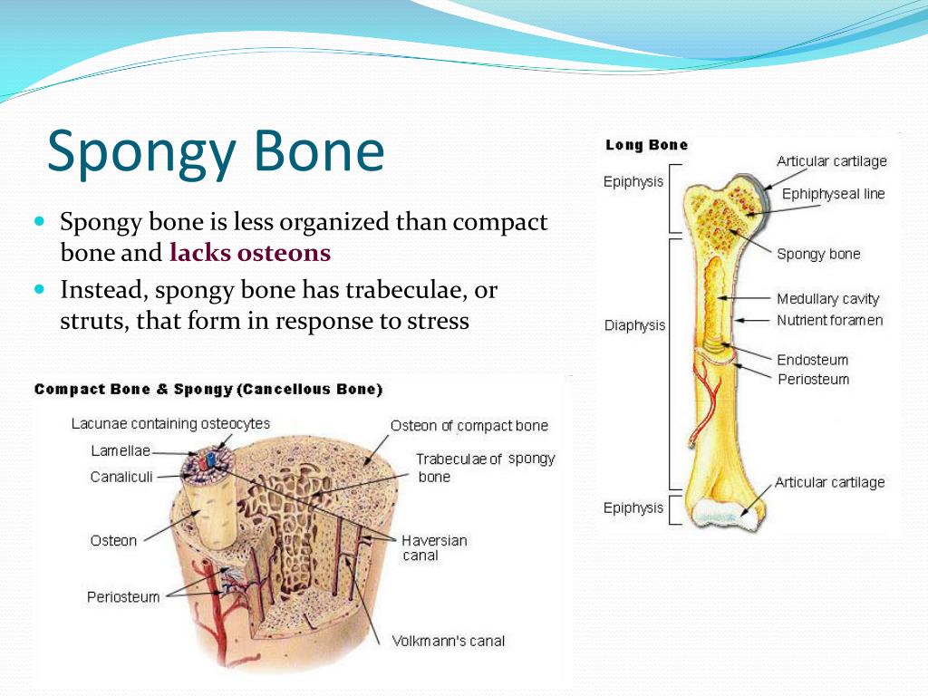
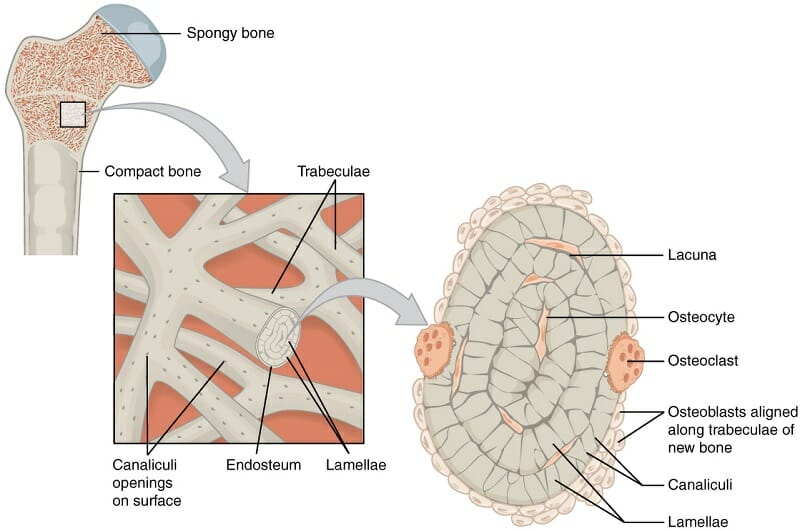
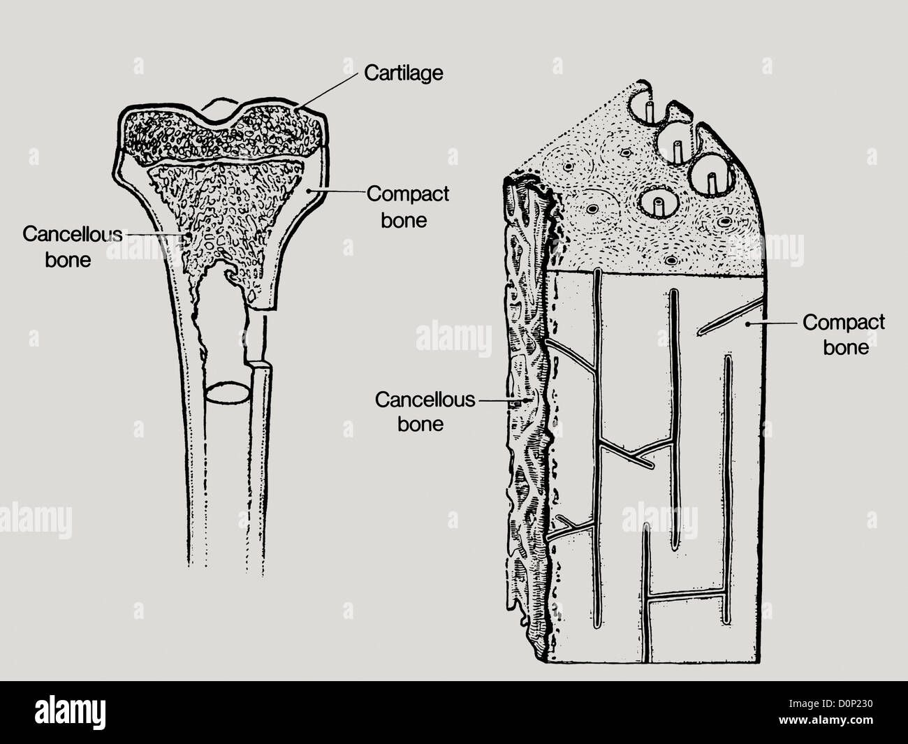
Spongy Bone Drawing - Spongy bone is usually located at the ends of the long bones (the epiphyses), with the harder compact bone surrounding it. Locate and name the major parts of a long bone. Web red marrow fills the spaces in the spongy bone. The spaces within the spongy bone are filled with red bone marrow containing the stem cells for blood cell production (the process of hematopoesis). It is surrounded by a layer of dense compact bone, creating a strong but lightweight bone structure. Web in this article you will learn on spongy bone histology with slide images and labelled diagram. Web histology of spongy bone how to draw spongy bone histologyhow to draw histology journalstep by step drawing of spongy bone with explanation spongy bone/cance. Web flat bones, like those of the cranium, consist of a layer of diploë (spongy bone), lined on either side by a layer of compact bone. Spongy bone makes up the interior of most bones and is located deep to the compact bone. In long bones, spongy bone forms the interior of the epiphyses; In long bones, spongy bone forms the interior of the epiphyses; Compact bone is the tissue that forms the surface of bones. Web flat bones, like those of the cranium, consist of a layer of diploë (spongy bone), lined on either side by a layer of compact bone. Osteocytes can be seen in layers in adult spongy bone. Web red. Web spongy bone, also called cancellous or trabecular bone, is a porous tissue that forms the interior of bones, especially the epiphysis and metaphysis of long bones. A cross section of the bone shows compact bone and blood vessels in the bone marrow. Spongy bone is supplied by fewer and larger vessels than compact bone. While it is not as. Web histology of spongy bone how to draw spongy bone histologyhow to draw histology journalstep by step drawing of spongy bone with explanation spongy bone/cance. Web about press copyright contact us creators advertise developers terms privacy policy & safety how youtube works test new features nfl sunday ticket press copyright. Drawing shows spongy bone, red marrow, and yellow marrow. From. Spongy bone is formed by an anastomosing network of trabeculae that form interconnecting spaces containing bone marrow. Web spongy bone provides balance to the dense and heavy compact bone by making bones lighter so that muscles can move them more easily. Web spongy bone, also called cancellous or trabecular bone, is a porous tissue that forms the interior of bones,. Web these irregularly shaped bones have a thin cortical shell which surrounds spongy (cancellous or trabecular) bone (#1 and #2). The spaces within the spongy bone are filled with red bone marrow containing the stem cells for blood cell production (the process of hematopoesis). In long bones, spongy bone forms the interior of the epiphyses; Osteocytes can be seen in. Spongy bone is composed of trabeculae that contain the osteocytes. Compact bone is the tissue that forms the surface of bones. It is surrounded by a layer of dense compact bone, creating a strong but lightweight bone structure. Also shown are red blood cells, white blood cells, platelets, and a blood stem cell. The two layers of compact bone and. Each epiphysis meets the diaphysis at the metaphysis, the narrow area that contains the epiphyseal plate (growth plate), a layer of hyaline (transparent) cartilage in a growing bone. Best spongy bone histology slide images The spaces within the spongy bone are filled with red bone marrow containing the stem cells for blood cell production (the process of hematopoesis). Web both. In long bones, spongy bone forms the interior of the epiphyses; Best spongy bone histology slide images It is highly vascularized and contains red bone marrow. Web these irregularly shaped bones have a thin cortical shell which surrounds spongy (cancellous or trabecular) bone (#1 and #2). A cross section of the bone shows compact bone and blood vessels in the. Distinguish between compact and spongy bone. Web both the epiphyses and metaphyses have a thin cortical layer of compact bone that is filled with a porous bone arrangement called spongy bone. Osteocytes can be seen in layers in adult spongy bone. Each epiphysis meets the diaphysis at the metaphysis, the narrow area that contains the epiphyseal plate (growth plate), a. Web flat bones, like those of the cranium, consist of a layer of diploë (spongy bone), lined on either side by a layer of compact bone. Web diagram of spongy bone. Spongy bone makes up the interior of most bones and is located deep to the compact bone. Drawing shows spongy bone, red marrow, and yellow marrow. Spongy bone is. Locate and name the major parts of a long bone. In addition, the spaces in some spongy bones contain red bone marrow, protected by the trabeculae, where hematopoiesis occurs. Web anatomy of the bone; Web in this article you will learn on spongy bone histology with slide images and labelled diagram. Web diagram of spongy bone. Web histology of spongy bone how to draw spongy bone histologyhow to draw histology journalstep by step drawing of spongy bone with explanation spongy bone/cance. Compact bone is the tissue that forms the surface of bones. Describe the functions of various parts of a bone. Web spongy bone, also called cancellous or trabecular bone, is a porous tissue that forms the interior of bones, especially the epiphysis and metaphysis of long bones. This produces a light, porous bone, that is strong. The two layers of compact bone and the interior spongy bone work together to. Web spongy bone is on the interior of a bone and consists of slender fibers and lamellae—layers of bony tissue—that join to form a reticular structure. The spaces within the spongy bone are filled with red bone marrow containing the stem cells for blood cell production (the process of hematopoesis). The diaphysis (shaft) consists of compact bone surrounding the. Web these irregularly shaped bones have a thin cortical shell which surrounds spongy (cancellous or trabecular) bone (#1 and #2). Web spongy bone is the tissue that makes up the interior of bones;
Histology of Spongy Bone YouTube

14 How to Draw Spongy Bone/Histology/Exams Prep YouTube

Histology Glossary Histology Spongy Bone Draw It to Know It

Compact And Spongy Bone Diagram

Structure of spongy bone

diagram of spongy bone

Spongy Bone (Cancellous Bone) Definition & Function Biology

Structure of Spongy Bone

Spongy Bone (Label) Diagram Quizlet

Compact Spongy Bone Diagram
Web Spongy Bone Contains Large Marrow Spaces Defined By Shelves And Spicules Of Bone.
The Two Layers Of Compact Bone And The Interior Spongy Bone Work Together To Protect The Internal Organs.
Drawing Shows Spongy Bone, Red Marrow, And Yellow Marrow.
Also Shown Are Red Blood Cells, White Blood Cells, Platelets, And A Blood Stem Cell.
Related Post: