Smooth Muscle Drawing
Smooth Muscle Drawing - Smooth muscle cells (orange with purple nuclei) are embedded in the. How to draw a muscle tissue | straight muscles | smooth muscles | cardiac muscleshello friends in this video i tell you about how to draw labelled dia. It is found throughout arteries and veins where it. Web smooth muscle cells, also known as muscle fibers, are generally arranged in bundles. And the longitudinal components with gap junctions. H&e and ihc staining were used to analyse the histopathology of. Intracellular actin and myosin filaments generate contractile forces by a sliding filament mechanism. Web from the smooth muscle histology slide, you might identify the following important histological features. Smooth muscle cells are a lot smaller than cardiac muscle cells, and they do not branch or connect end to end the way cardiac cells do. Search images from huge database containing over 1,250,000 drawings Web loose weight concept, woman with a regular normal body with an abdomen muscles drawing. In relaxed smooth muscle, the nuclei are elongated with rounded ends. Unlike cardiac and skeletal muscle cells, smooth muscle cells do not exhibit striations since their actin. Smooth muscle tissue is composed of sheets or strands of cells. Smooth muscle cells are a lot smaller. Search images from huge database containing over 1,250,000 drawings Web this type of smooth muscle is observed in the large airways to the lungs, in the large arteries, the arrector pili muscles associated with hair follicles, and the internal eye muscles which regulate light entry and lens shape. Web loose weight concept, woman with a regular normal body with an. Smooth muscle cells or fibres of the longitudinal section #2. Web smooth muscle cells, also known as muscle fibers, are generally arranged in bundles. Schematic drawing of a longitudinal section of a bundle of smooth muscle cells. Lobata effect on the proliferation and inflammation of vascular smooth muscle in as and the potential mechanism were investigated. H&e and ihc staining. Smooth muscle will have many “empty” cells in which the nucleus is out of the plane of section. Like striated muscle, smooth muscle can tense and relax. Smooth muscle cells or fibres of the longitudinal section #2. It is found throughout arteries and veins where it. The nucleus of smooth muscle cells or fibres of the longitudinal section #3. It is found throughout arteries and veins where it. Wall of organs like the stomach, oesophagus and intestine. Web smooth muscle is one of three types of muscle tissue, alongside cardiac and skeletal muscle. Smooth muscle tissue is found around organs in the digestive, respiratory. Contractile filaments are not arranged into sarcomeres, thus giving it a nonstriated (smooth. Smooth muscle cells or fibres of the longitudinal section #2. Web free download 43 best quality smooth muscle drawing at getdrawings. Web smooth muscle cells, also known as muscle fibers, are generally arranged in bundles. Smooth muscle cells range in size from 0.2 micrometers to ten micrometers in diameter and from fifty micrometers to. It is the pen diagram of. Smooth muscle cells (orange with purple nuclei) are embedded in the. And the longitudinal components with gap junctions. It is in the stomach and intestines where it helps with digestion and nutrient collection. How to draw smooth muscle cell diagram | how to draw smooth muscle cell easilyhello friends in this video i tell you about how to draw smooth. Smooth muscle tissue is found around organs in the digestive, respiratory. Intracellular actin and myosin filaments generate contractile forces by a sliding filament mechanism. Smooth muscle cells are a lot smaller than cardiac muscle cells, and they do not branch or connect end to end the way cardiac cells do. It is in the stomach and intestines where it helps. Contractile filaments are not arranged into sarcomeres, thus giving it a nonstriated (smooth. Lymphatic system, vintage engraved illustration. Connective tissue surrounds the smooth muscle fibers or cells with fibroblasts #4. How to draw smooth muscle cell diagram | how to draw smooth muscle cell easilyhello friends in this video i tell you about how to draw smooth muscle c. The. Smooth muscle cells range in size from 0.2 micrometers to ten micrometers in diameter and from fifty micrometers to. Web drawing histological diagram of transverse section of smooth muscle.useful for all medical students(mbbs,bhms,bams,bpt,ahs,nursing)drawn by using h & e penci. Web smooth muscle cells, also known as muscle fibers, are generally arranged in bundles. Smooth muscle tissue is composed of sheets. Smooth muscle cells are a lot smaller than cardiac muscle cells, and they do not branch or connect end to end the way cardiac cells do. And the longitudinal components with gap junctions. It is the pen diagram of skeletal, smooth and cardiac muscle for class 10, 11 and 12. Web smooth muscle is ideally seen in the wall of hollow visceral organs, including different parts of the compound stomach, simple stomach, various parts of the intestine, urinary bladder, and uterus, #2. Web from the smooth muscle histology slide, you might identify the following important histological features. Physiologically divided into single unit and multi unit fibers. Contractile filaments are not arranged into sarcomeres, thus giving it a nonstriated (smooth. You will also find the smooth muscle layers in these organs or structures that form various narrow tubes; H&e and ihc staining were used to analyse the histopathology of. Connective tissue surrounds the smooth muscle fibers or cells with fibroblasts #4. How to draw smooth muscle cell diagram | how to draw smooth muscle cell easilyhello friends in this video i tell you about how to draw smooth muscle c. Smooth muscle cells (orange with purple nuclei) are embedded in the. Web male nod colonic smooth muscle exhibited decreased h3k27ac levels, not female, whereas female nod colonic smooth muscle demonstrated diminished enrichment of h3ac at the gper promoter, contrary to male nod. Lobata effect on the proliferation and inflammation of vascular smooth muscle in as and the potential mechanism were investigated. Search images from huge database containing over 1,250,000 drawings Smooth muscle tissue, unlike striated muscle, contracts slowly and automatically.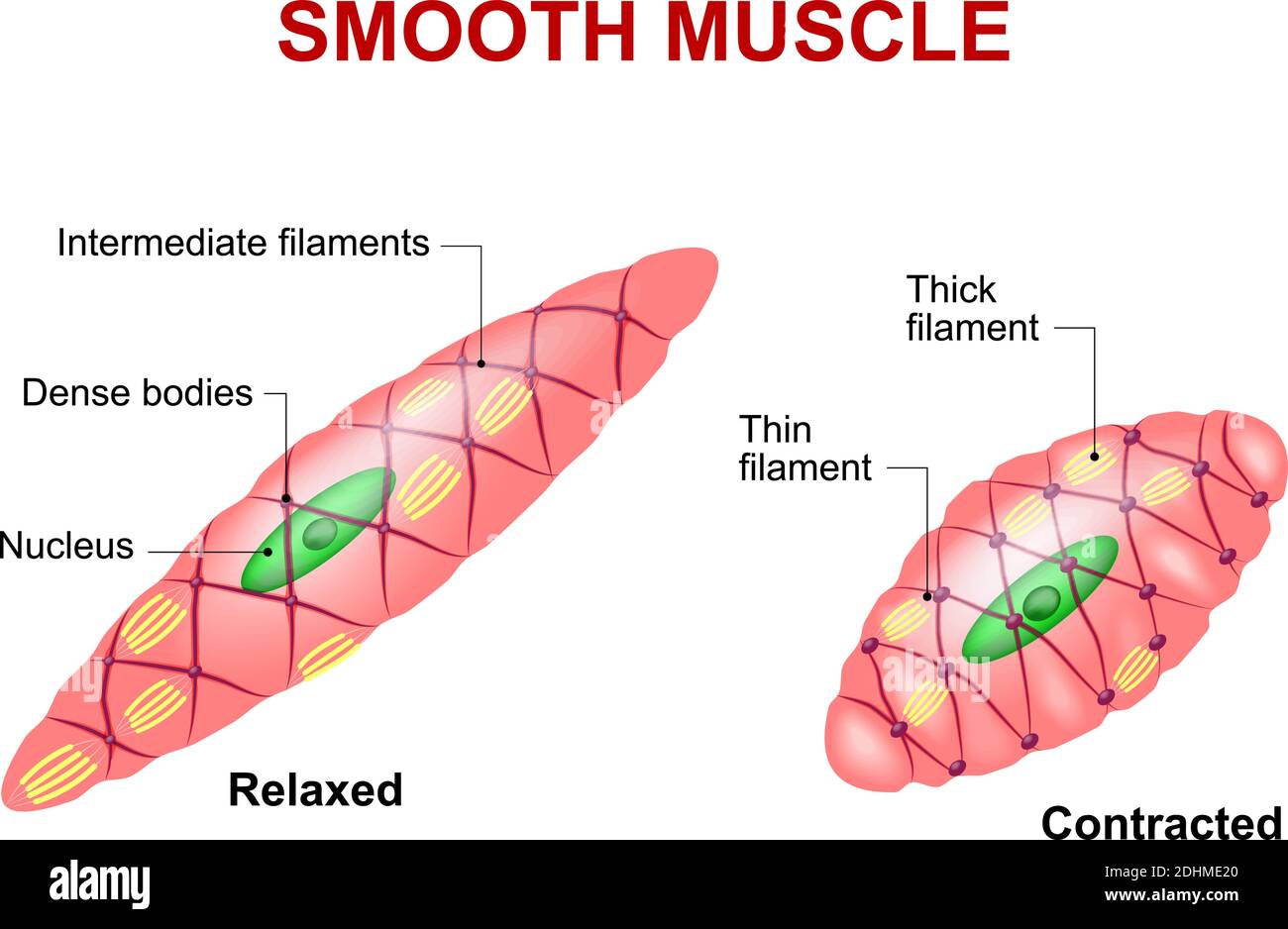
Smooth muscle tissue. Anatomy of a relaxed and contracted smooth muscle

10.8 Smooth Muscle Douglas College Human Anatomy and Physiology I
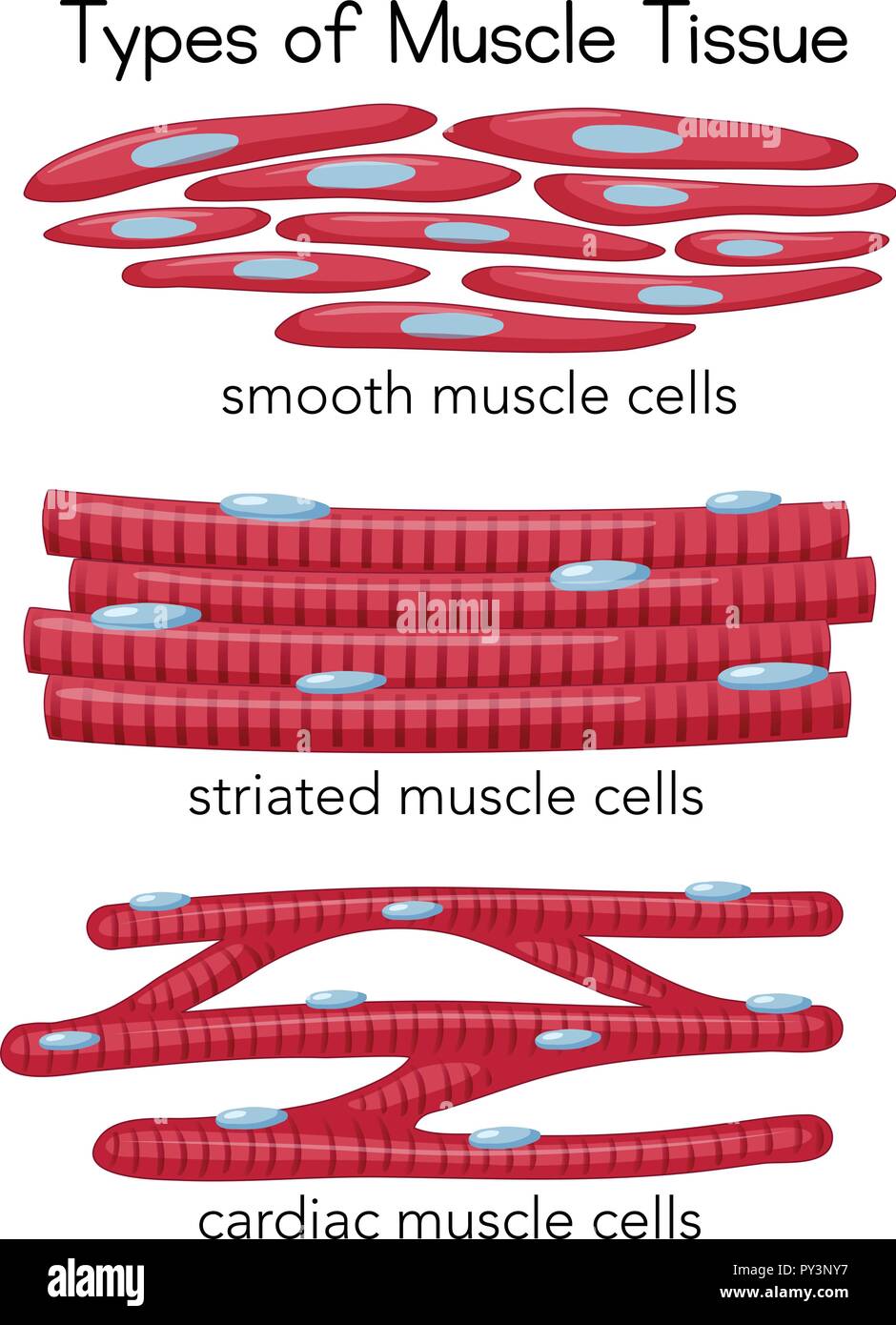
Smooth Muscle Diagram Drawing Notez On Nursing.... Tissues Muscle

Smooth Muscle Diagram Drawing Notez On Nursing.... Tissues Muscle
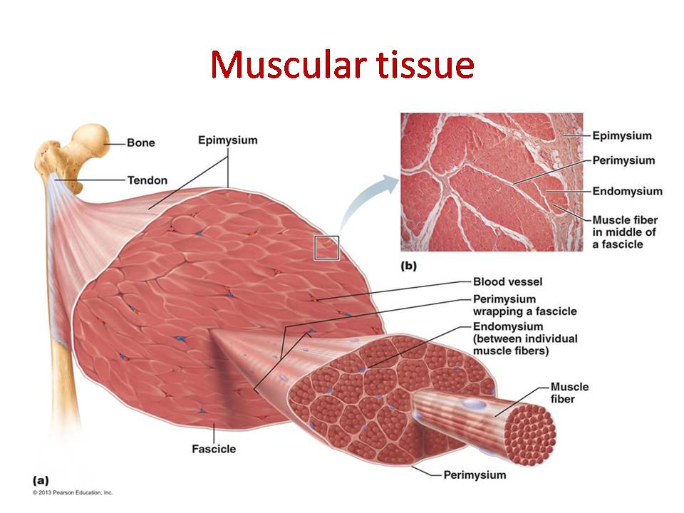
Smooth Muscle Diagram / Muscle cell diagram Smooth muscle anatomy

Smooth Muscle Diagram Labeled Muscles Of The Body Diagrams, How
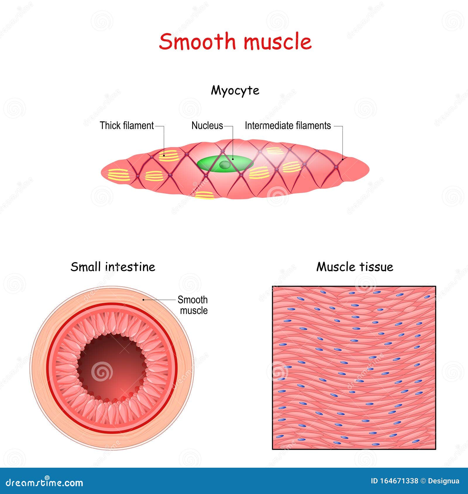
Smooth Muscle Anatomy

Smooth Muscle Diagram Smooth Muscle Hd Stock Images Shutterstock
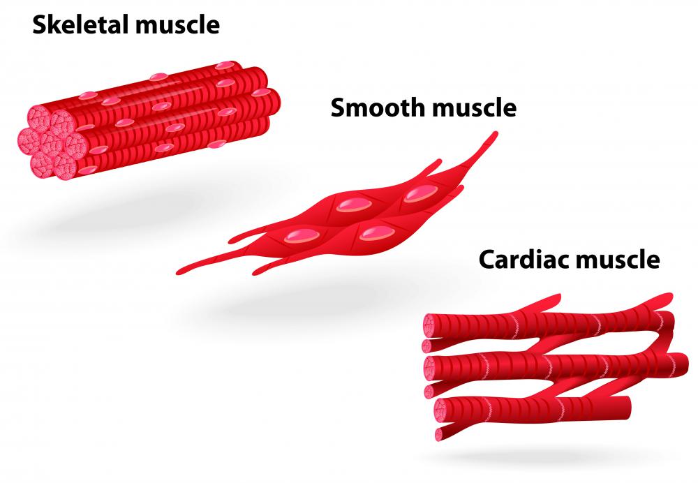
What is a Smooth Muscle Contraction? (with pictures)

Smooth Muscle Tissue Diagram Drawing ezildaricci
Smooth Muscle Tissue Is Composed Of Sheets Or Strands Of Cells.
Smooth Muscle, Muscle That Shows No Cross Stripes Under Microscopic Magnification.
Web In This Video I Have Shown The Simplest Way Of Drawing Muscle Drawing.
Web Drawing Histological Diagram Of Transverse Section Of Smooth Muscle.useful For All Medical Students(Mbbs,Bhms,Bams,Bpt,Ahs,Nursing)Drawn By Using H & E Penci.
Related Post: