Pineal Cyst Size Chart
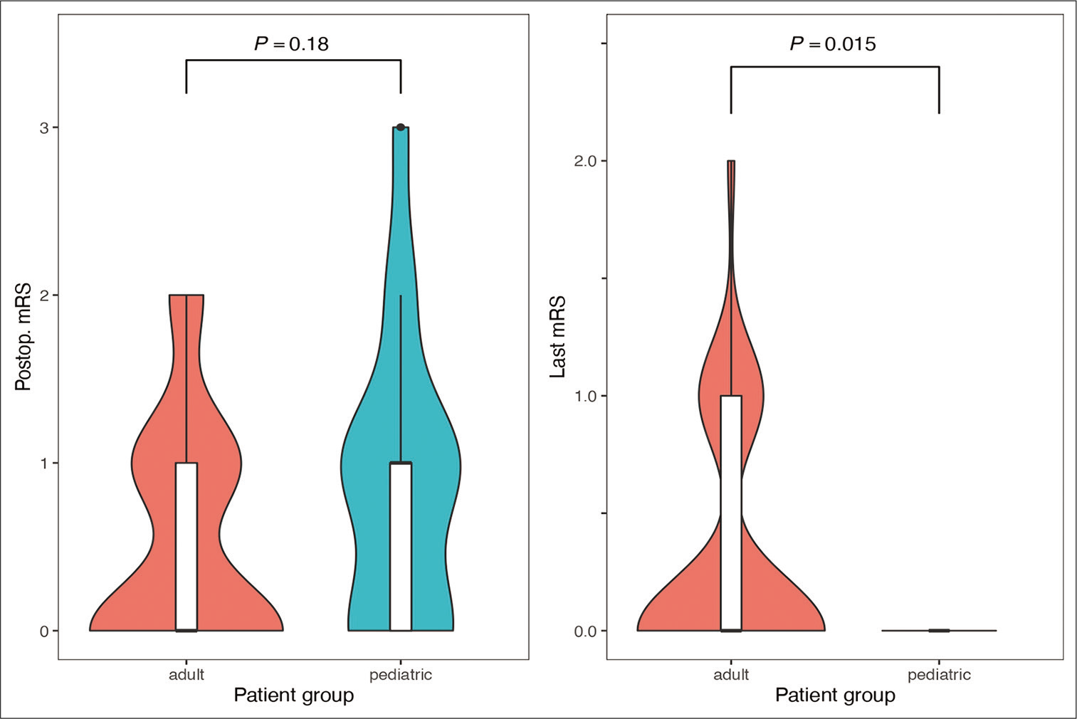
Pineal Cyst Size Chart - Web scatter chart showing patients with pineal cyst progression and regression by age and cyst diameter full size image analyzing patients according to cyst size change, we found a significant difference in the mean age between the pc progression group and pc regression group ( p = 0.01). Web although most pineal cysts are small, large pineal cysts can cause a variety of symptoms such as: In most cases, no treatment is necessary for a pineal gland cyst. Web a total of 294 patients with surgically managed nhspc were identified. Web pineal gland cysts are common. As many as 2 percent of healthy adults develop this kind of cyst. Web accurate diagnosis is critical in determining the nature of a pineal cyst and the appropriate treatment, if necessary. [ 4, 15, 29] symptoms such as headache, hydrocephalus and visual deficiency have been described in classic cases. Web we aimed to identify predictive factors of pineal cyst volume change and surgical intervention by performing retrospective chart review of 98 patients between 2005 and 2018 diagnosed with pineal cysts gleaned from our neurosurgery clinical databases. Confirm the presence of a pineal cyst. Imaging plays a vital role in the diagnostic process by allowing healthcare professionals to: Mean age was 29 (range: Cysts can form in all parts of the body, including the brain. Confirm the presence of a pineal cyst. Finely fibrillary glial tissue often containing hemosiderin. Web scatter chart showing patients with pineal cyst progression and regression by age and cyst diameter full size image analyzing patients according to cyst size change, we found a significant difference in the mean age between the pc progression group and pc regression group ( p = 0.01). Web pineal cysts can be categorised on mr imaging as either simple. [ 4, 15, 29] symptoms such as headache, hydrocephalus and visual deficiency have been described in classic cases. Web pineal gland cysts are common. 1 since the advent of mr brain imaging, the incidence of pineal cysts has been reported to vary from 0.58% to 10.8% in large consecutive brain mr imaging studies. Imaging plays a vital role in the. [ 4, 15, 29] symptoms such as headache, hydrocephalus and visual deficiency have been described in classic cases. Web we aimed to identify predictive factors of pineal cyst volume change and surgical intervention by performing retrospective chart review of 98 patients between 2005 and 2018 diagnosed with pineal cysts gleaned from our neurosurgery clinical databases. Less, however, is known about. Web pineal cysts are composed of three concentric layers: Web symptomatic cysts vary in size from 7 mm to 45 mm, whereas asymptomatic cysts are usually less than 10 mm in diameter, although a relationship between the cyst size and the onset of symptoms has been proved to be irrelevant in many cases. Finely fibrillary glial tissue often containing hemosiderin.. Web pineal cyst (pc) is a relatively common true cyst in the pineal gland. Confirm the presence of a pineal cyst. Finely fibrillary glial tissue often containing hemosiderin. Less, however, is known about the prevalence and appearance of pineal cysts in children. Web accurate diagnosis is critical in determining the nature of a pineal cyst and the appropriate treatment, if. Less, however, is known about the prevalence and appearance of pineal cysts in children. [ 4, 15, 29] symptoms such as headache, hydrocephalus and visual deficiency have been described in classic cases. Web although most pineal cysts are small, large pineal cysts can cause a variety of symptoms such as: Web benign pineal cysts are usually incidental findings on magnetic. Assess the size and location of the cyst. In most cases, no treatment is necessary for a pineal gland cyst. Rarely does a pineal gland cyst cause headaches or any other symptoms. Less, however, is known about the prevalence and appearance of pineal cysts in children. Mean age was 29 (range: Confirm the presence of a pineal cyst. The maximal dimension of the cyst did not change in 24 (75%) of 32 patients. Web maximal cyst dimensions ranged from 0.5 to 2.2 cm. Web pineal cyst (pc) is a relatively common true cyst in the pineal gland. Web pineal cysts are composed of three concentric layers: 3 simple pineal cysts are unilocular, often with a smooth, thin wall (which may or may not enhance) and can range in size from <<strong>5 mm to</strong> >25 mm in maximum diameter ( figure 1 ). Web although most pineal cysts are small, large pineal cysts can cause a variety of symptoms such as: Pineal parenchyma with or without calcification.. Web maximal cyst dimensions ranged from 0.5 to 2.2 cm. Web pineal cyst (pc) is a relatively common true cyst in the pineal gland. Mean age was 29 (range: Web scatter chart showing patients with pineal cyst progression and regression by age and cyst diameter full size image analyzing patients according to cyst size change, we found a significant difference in the mean age between the pc progression group and pc regression group ( p = 0.01). 1 since the advent of mr brain imaging, the incidence of pineal cysts has been reported to vary from 0.58% to 10.8% in large consecutive brain mr imaging studies. Web we aimed to identify predictive factors of pineal cyst volume change and surgical intervention by performing retrospective chart review of 98 patients between 2005 and 2018 diagnosed with pineal cysts gleaned from our neurosurgery clinical databases. Web benign pineal cysts are usually incidental findings on magnetic resonance imaging (mri). Web although most pineal cysts are small, large pineal cysts can cause a variety of symptoms such as: Imaging plays a vital role in the diagnostic process by allowing healthcare professionals to: Web pineal gland cysts are common. Web accurate diagnosis is critical in determining the nature of a pineal cyst and the appropriate treatment, if necessary. Rarely does a pineal gland cyst cause headaches or any other symptoms. The maximal dimension of the cyst did not change in 24 (75%) of 32 patients. Web symptomatic cysts vary in size from 7 mm to 45 mm, whereas asymptomatic cysts are usually less than 10 mm in diameter, although a relationship between the cyst size and the onset of symptoms has been proved to be irrelevant in many cases. In most cases, no treatment is necessary for a pineal gland cyst. 3 simple pineal cysts are unilocular, often with a smooth, thin wall (which may or may not enhance) and can range in size from <<strong>5 mm to</strong> >25 mm in maximum diameter ( figure 1 ).
Pineal cysts diameters across the surgical criteria groups (a

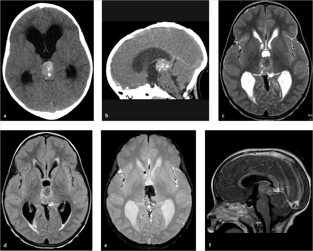
Pineal Cyst Simulating Pinealoblastoma in 11 Children With

Pineal cyst growth during followup, from 1 × 1.5 cm (a) to 1.5 × 1.9

Pineal Cyst Simulating Pinealoblastoma in 11 Children With

Scatter chart showing patients with pineal cyst progression and

Pineal Cyst Size Chart

11 Simple Pineal Cyst Radiology Key

Pineal Cyst Size Chart

Pineal Cyst Size Chart

Surgical Neurology International
As Many As 2 Percent Of Healthy Adults Develop This Kind Of Cyst.
Web Pineal Cysts Can Be Categorised On Mr Imaging As Either Simple Or Atypical.
Web A Total Of 294 Patients With Surgically Managed Nhspc Were Identified.
Finely Fibrillary Glial Tissue Often Containing Hemosiderin.
Related Post: