Nerve Chart Spine
Nerve Chart Spine - It is important to mention that after the spinal nerves exit from the spine, they join together to form four paired clusters of. For the most part, the spinal nerves exit the vertebral canal through the intervertebral foramen below their corresponding vertebra. Numbers indicate the types of nerve fibers: Spinal cord and spinal nerve roots. L2, l3 and l4 spinal nerves provide sensation to the front part of your thigh and inner side of your lower leg. Spinal nerves emerge from the spinal cord and reorganize through plexuses, which then give rise to systemic nerves. On the chart below you will see 4 columns (vertebral level, nerve root, innervation, and possible symptoms). These nerves also control movements of the hip and knee muscles. Your spinal cord is a column of nerves that travels through your spinal canal. The cord extends from your skull to your lower back. These nerves carry messages between your brain and muscles. Web to visualize the spinal nerves, a comprehensive chart is needed that details the anatomy and physiology of the nervous system. Web spinal nerves are all mixed nerves with both sensory and motor fibers. Web l1 spinal nerve provides sensation to the groin and genital regions and may contribute to the. These nerves also control movements of the hip and knee muscles. Web to visualize the spinal nerves, a comprehensive chart is needed that details the anatomy and physiology of the nervous system. Web spinal nerves are all mixed nerves with both sensory and motor fibers. Each of these nerves branches out from the spinal cord, dividing and subdividing to form. Thoracic spinal nerves are not part of any plexus, but give rise to the intercostal nerves directly. The dorsal root is the afferent sensory root and carries sensory information to the brain. You have 31 spinal nerves and 30 dermatomes. Web dermatomes are areas of skin that are connected to a single spinal nerve. Spinal cord and spinal nerve roots. These nerves also control movements of the hip and knee muscles. Web cervical radiculopathy (also known as “pinched nerve”) is a condition that results in radiating pain, weakness and/or numbness caused by compression of any of the nerve roots in your neck. The back is the body region between the neck and the gluteal regions. Most cases of cervical radiculopathy. Web the spine’s four sections, from top to bottom, are the cervical (neck), thoracic (abdomen,) lumbar (lower back), and sacral (toward tailbone). If this complex system is damaged, nerve signals can go awry, causing intense pain. Spinal nerves emerge from the spinal cord and reorganize through plexuses, which then give rise to systemic nerves. Web to visualize the spinal nerves,. These nerves also control movements of the hip and knee muscles. This diagram indicates the formation of a typical spinal nerve from the dorsal and ventral roots. Web cervical radiculopathy (also known as “pinched nerve”) is a condition that results in radiating pain, weakness and/or numbness caused by compression of any of the nerve roots in your neck. Web the. If this complex system is damaged, nerve signals can go awry, causing intense pain. Web below is a chart that outlines the main functions of each of the spine nerve roots: Web how to use the spinal nerve chart: The cord extends from your skull to your lower back. The back is the body region between the neck and the. Your spinal cord is a column of nerves that travels through your spinal canal. Eight cervical spinal nerve pairs, 12 thoracic pairs , five lumbar pairs, five sacral pairs, and one coccygeal. For the most part, the spinal nerves exit the vertebral canal through the intervertebral foramen below their corresponding vertebra. These nerves also control movements of the hip and. Most cases of cervical radiculopathy go away with nonsurgical treatment. Spinal cord and spinal nerve roots. Spinal nerves emerge from the spinal cord and reorganize through plexuses, which then give rise to systemic nerves. The dorsal root is the afferent sensory root and carries sensory information to the brain. Web dermatomes are areas of skin that are connected to a. Thomas scioscia, md , orthopedic surgeon. Throughout the spine, intervertebral discs made of. Each nerve forms from nerve fibers, known as fila radicularia, extending from the posterior (dorsal) and anterior (ventral) roots of the spinal cord. Your spinal cord is a column of nerves that travels through your spinal canal. You have 31 spinal nerves and 30 dermatomes. Eight cervical spinal nerve pairs, 12 thoracic pairs , five lumbar pairs, five sacral pairs, and one coccygeal. These nerves also control movements of the hip and knee muscles. It is important to mention that after the spinal nerves exit from the spine, they join together to form four paired clusters of. These nerves carry messages between your brain and muscles. You have 31 spinal nerves and 30 dermatomes. Web spinal nerves are mixed nerves that interact directly with the spinal cord to modulate motor and sensory information from the body’s periphery. Ralph rashbaum, md, orthopedic surgeon. L2, l3 and l4 spinal nerves provide sensation to the front part of your thigh and inner side of your lower leg. Web to visualize the spinal nerves, a comprehensive chart is needed that details the anatomy and physiology of the nervous system. This diagram indicates the formation of a typical spinal nerve from the dorsal and ventral roots. Web spine nerves anatomy, diagram & function | body maps. Web spinal nerves are all mixed nerves with both sensory and motor fibers. L2, l3, and l4 spinal nerves provide sensation to the front part of the thigh and inner side of the lower leg. The cord extends from your skull to your lower back. Throughout the spine, intervertebral discs made of. Numbers indicate the types of nerve fibers: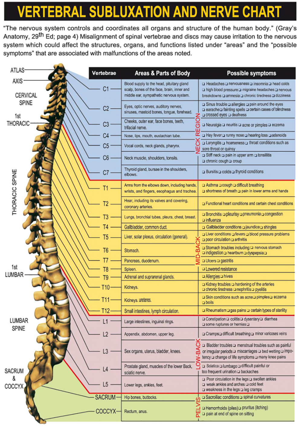
Anatomical Pain Chart

Spinal nerve function Medicina Pinterest Anatomía, Salud y Acupuntura
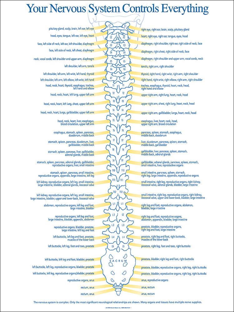
Chiropractic Spinal Nerve Chart Nerve Function Chart
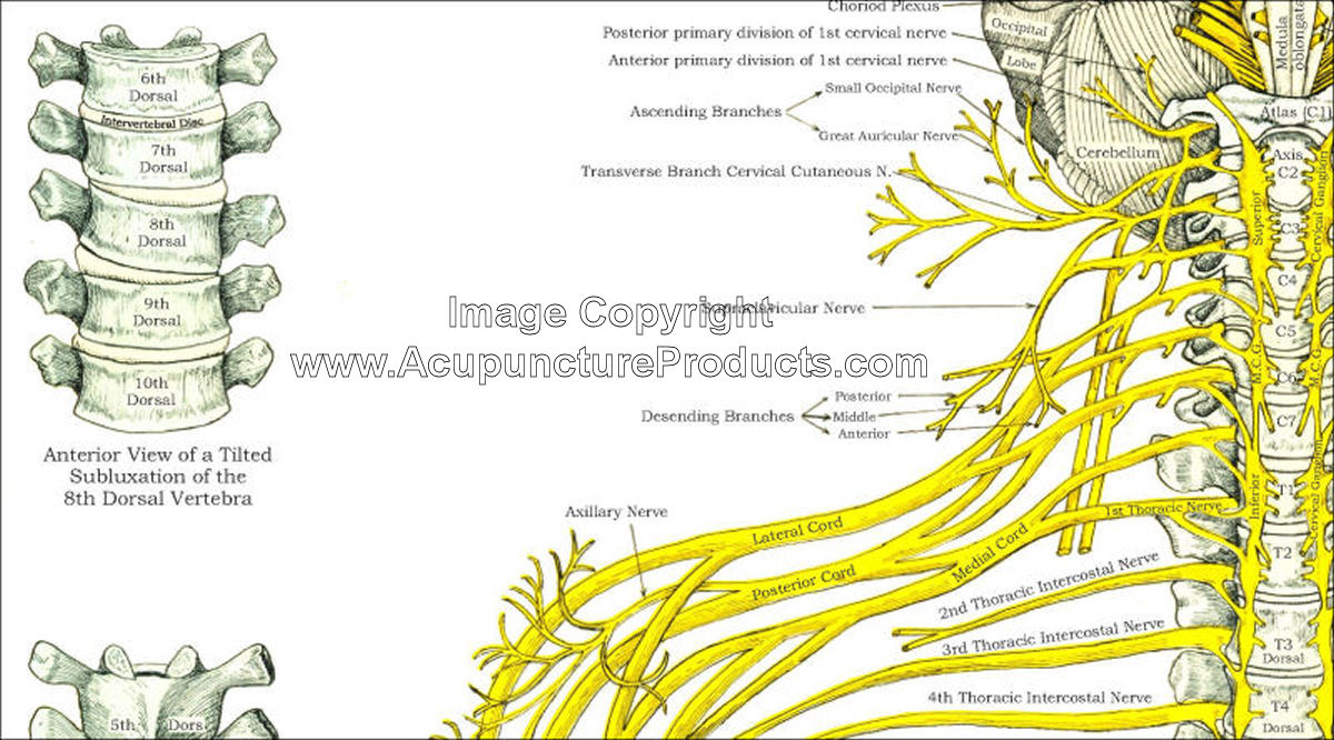
Chart Of Spine And Nerves
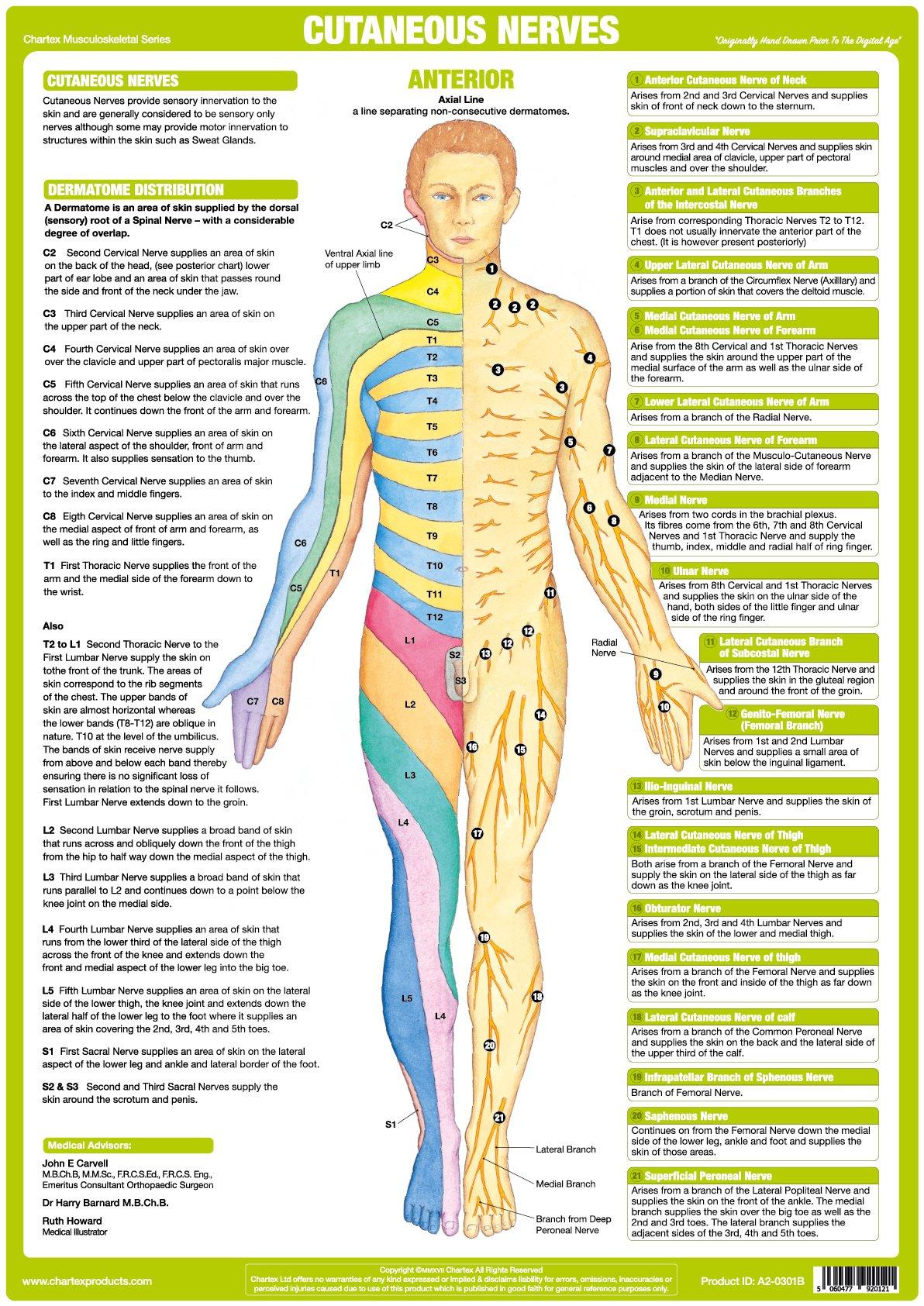
Nervous System Anatomy Posters Set of 6

Spinal Nerve Function Anatomical Chart Anatomy Models and Anatomical

Printable Spinal Nerve Chart Free Printable Calendar

Spinal Nerve Function , Vintage , Spinal Nerve Function Chart, Root

Printable Spinal Nerve Chart
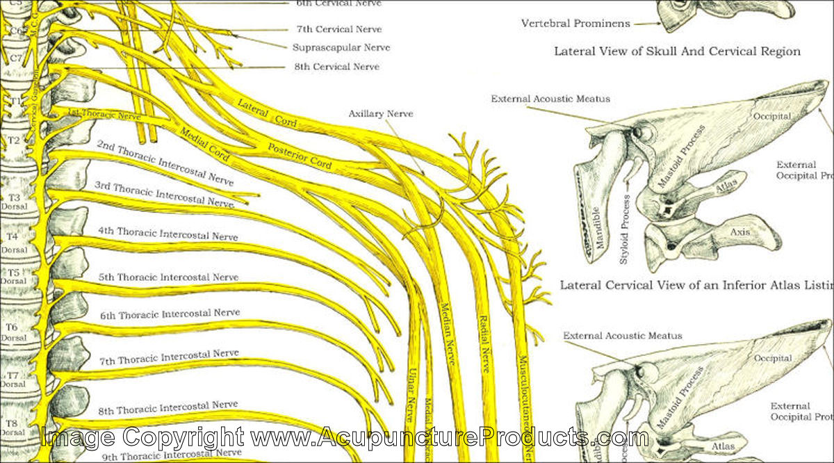
Vertebral Subluxation Spinal Nerves Chart
If This Complex System Is Damaged, Nerve Signals Can Go Awry, Causing Intense Pain.
Web Cervical Radiculopathy (Also Known As “Pinched Nerve”) Is A Condition That Results In Radiating Pain, Weakness And/Or Numbness Caused By Compression Of Any Of The Nerve Roots In Your Neck.
Web How To Use The Spinal Nerve Chart:
Web These Relay Motor (Movement), Sensory (Sensation), And Autonomic (Involuntary Functions) Signals Between The Spinal Cord And Other Parts Of The Body.
Related Post: