Lumbar Spine Nerve Chart
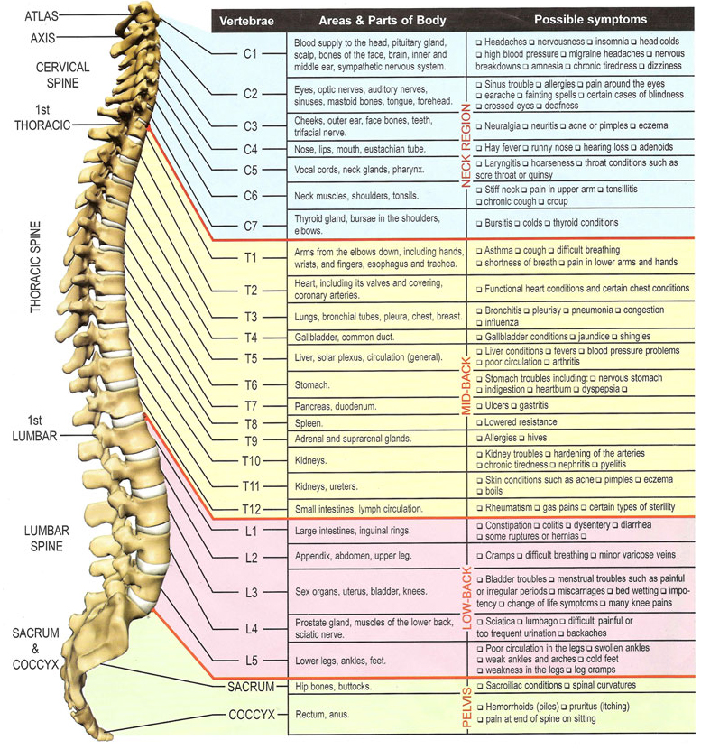
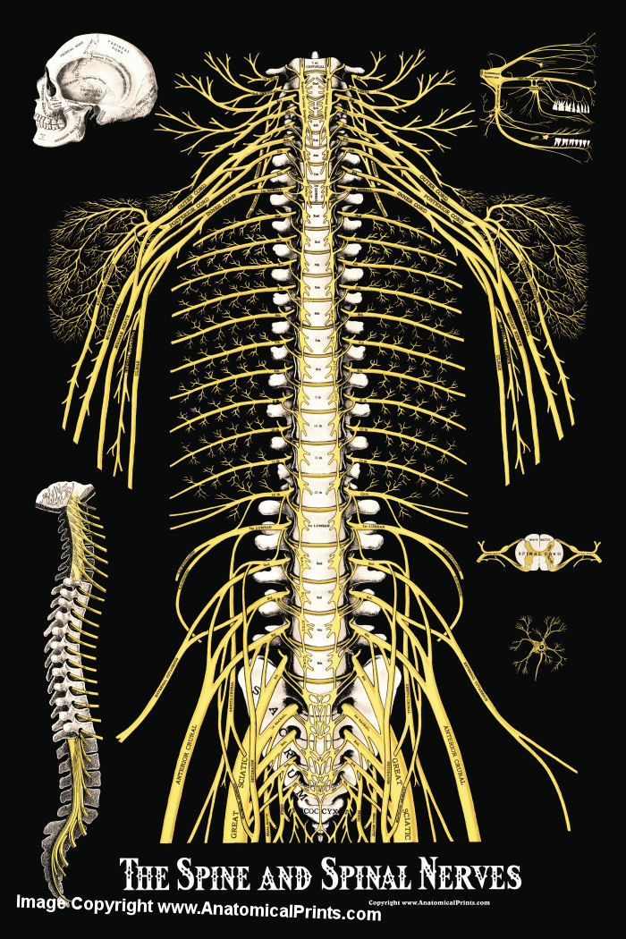
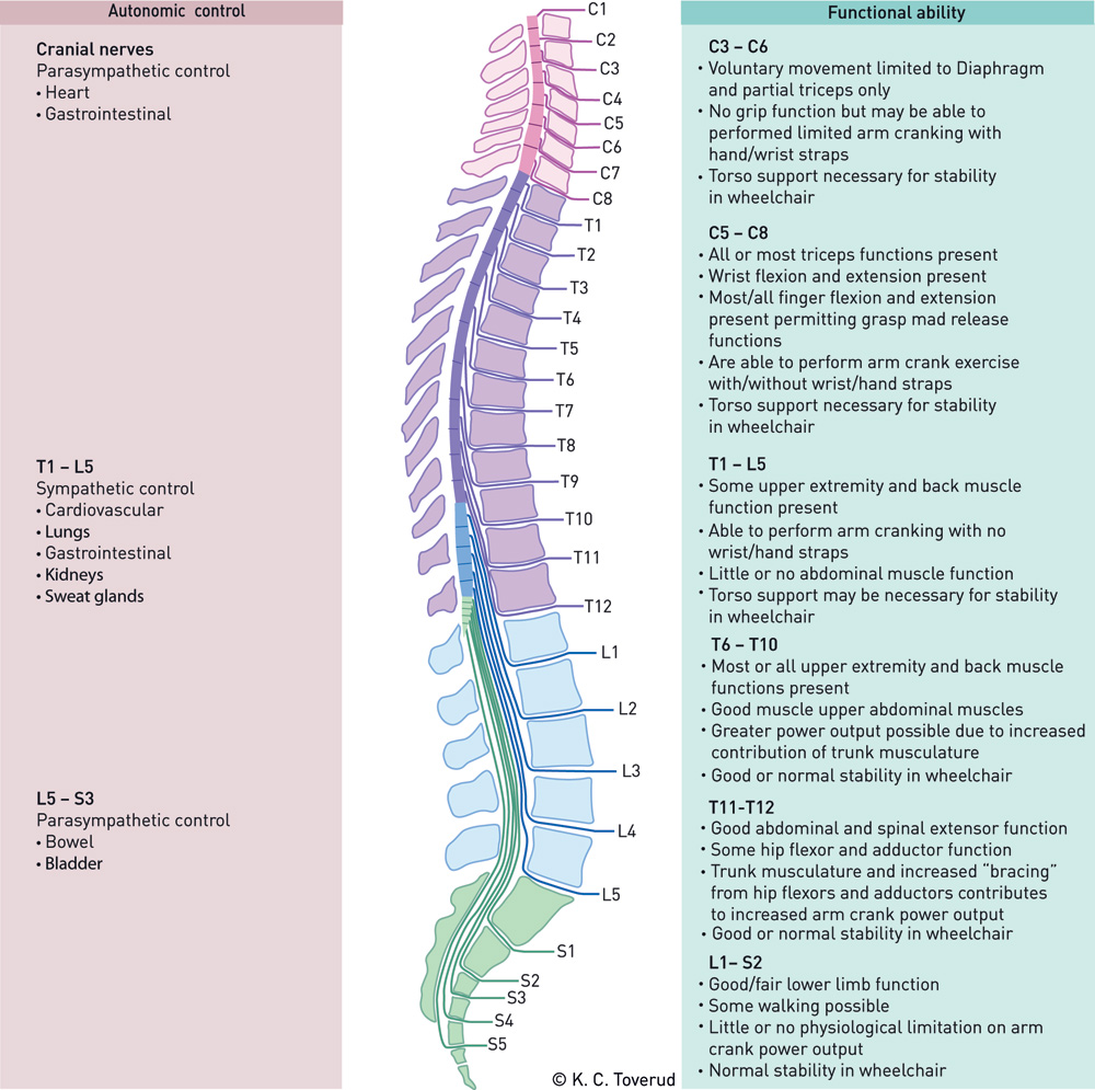
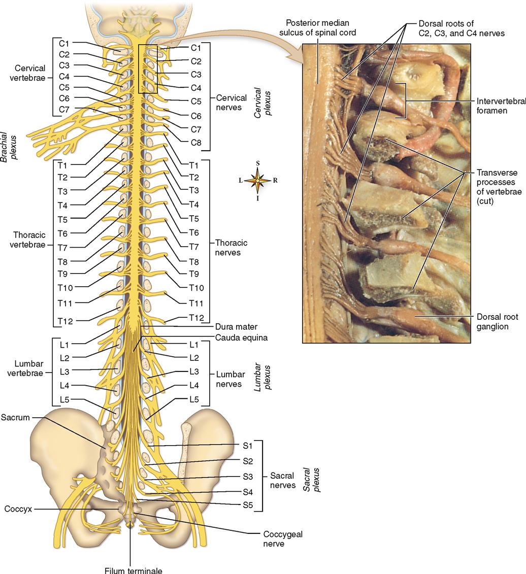
Lumbar Spine Nerve Chart - Web below is a chart that outlines the main functions of each of the spine nerve roots: While the nerves branch directly from the spinal cord and the central nervous system, the spinal nerves classify as a part of the peripheral nervous. Web the plexus is formed by the anterior rami (divisions) of the lumbar spinal nerves l1, l2, l3 and l4. It is important to mention that after the spinal nerves exit from the spine, they join together to form four paired clusters of. Web the relevant anatomy of the innervation of the musculature of the back by the spinal nerves is centered around the lumbar spinal nerves, peripheral nerves of the lumbar plexus, spinal cord, and lumbar vertebral column. For most spinal segments, the nerve roots run through the bony canal, and at each level a pair of nerve roots exits from the spine. The spine’s four sections, from top to bottom, are the cervical (neck), thoracic (abdomen,) lumbar (lower back), and. Web the lumbar nerves are five spinal nerves which arise from either side of the spinal cord below the thoracic spinal cord and above the sacral spinal cord. Giving your body structure (shape). Allowing you to be flexible and move. In general, the spinal cord consists of gray and white matter. While the nerves branch directly from the spinal cord and the central nervous system, the spinal nerves classify as a part of the peripheral nervous. These images alone won't show problems with the spinal cord, muscles, nerves or disks. Web highest risk of iatrogenic nerve injury to lumbar plexus. These images show arthritis or broken bones. They come from the part of your spine that makes up your lower back. Web the plexus is formed by the anterior rami (divisions) of the lumbar spinal nerves l1, l2, l3 and l4. These nerves also control movements of the hip and knee muscles. Allowing you to be flexible and move. Web what does the spine do? Web the lumbar nerves are five spinal nerves which arise from either side of the spinal cord below the thoracic spinal cord and above the sacral spinal cord. These scans generate images that can reveal herniated disks or problems with bones, muscles, tissue, tendons, nerves, ligaments and blood vessels. These images alone won't show. L5 spinal nerve provides sensation to the outer side of your lower leg, the upper part of your foot and the space between your first and second toe. Eight pairs of cervical nerves designated c 1 to c 8, twelve pairs of thoracic nerves designated t 1 to t 12, five pairs of lumbar nerves designated l 1 to l. Web therefore, there are 12 pairs of thoracic spinal nerves, 5 pairs of lumbar spinal nerves, 5 pairs of sacral spinal nerves, and a coccygeal nerve. Web together, the brain and spinal cord make up the central nervous system. These images show arthritis or broken bones. While the nerves branch directly from the spinal cord and the central nervous system,. However, this complex structure also leaves the low back susceptible to injury and pain. Web 5 lumbar spinal nerves. Web below is a chart that outlines the main functions of each of the spine nerve roots: These nerves also control hip and knee muscle movements. The spine’s four sections, from top to bottom, are the cervical (neck), thoracic (abdomen,) lumbar. L2, l3, and l4 spinal nerves provide sensation to the front part of the thigh and inner side of the lower leg. These images show arthritis or broken bones. While the nerves branch directly from the spinal cord and the central nervous system, the spinal nerves classify as a part of the peripheral nervous. Web the l4 and l5 are. These nerves also control hip and knee muscle movements. Sensory nerves are nerves that receive sensory stimuli, telling us how something feels—whether it is hot, cold, or painful. Web l1 spinal nerve provides sensation to the groin and genital regions and may contribute to the movement of the hip muscles. Functional anatomy of the lumbar spine. These images show arthritis. Web the peripheral nerves include both motor nerves and sensory nerves: These nerves also control hip and knee muscle movements. Sensory nerves are nerves that receive sensory stimuli, telling us how something feels—whether it is hot, cold, or painful. Web l2, l3 and l4 spinal nerves provide sensation to the front part of your thigh and inner side of your. Web l2, l3 and l4 spinal nerves provide sensation to the front part of your thigh and inner side of your lower leg. The lumbar vertebrae, as a group, produce a lordotic curve [1] the intervertebral discs are responsible for the mobility without sacrificing the supportive strength of the vertebral column. The spine’s four sections, from top to bottom, are. These nerves also control movements of the hip and knee muscles. When viewed from the side, the lumbar spine has a concave lordotic curve that helps distribute weights and reduce the. Spinal nerves can be impacted by a variety of medical conditions, resulting in pain, weakness, or decreased sensation. Web the plexus is formed by the anterior rami (divisions) of the lumbar spinal nerves l1, l2, l3 and l4. It is important to mention that after the spinal nerves exit from the spine, they join together to form four paired clusters of. Web l1 spinal nerve provides sensation to the groin and genital regions and may contribute to the movement of the hip muscles. These images alone won't show problems with the spinal cord, muscles, nerves or disks. Web these relay motor (movement), sensory (sensation), and autonomic (involuntary functions) signals between the spinal cord and other parts of the body. Web the lumbar spinal nerves that branch off from the spinal cord and cauda equina to control movements and sensation in the legs. The spine’s four sections, from top to bottom, are the cervical (neck), thoracic (abdomen,) lumbar (lower back), and. L2, l3, and l4 spinal nerves provide sensation to the front part of the thigh and inner side of the lower leg. They come from the part of your spine that makes up your lower back. For most spinal segments, the nerve roots run through the bony canal, and at each level a pair of nerve roots exits from the spine. Web l2, l3 and l4 spinal nerves provide sensation to the front part of your thigh and inner side of your lower leg. L5 spinal nerve provides sensation to the outer side of your lower leg, the upper part of your foot and the space between your first and second toe. Giving your body structure (shape).
Spinal nerve function from Anatomy in Motion Spine health, Spinal

Lumbar Spinal Nerve Chart

Spinal Column Anatomy Illustration Anatomy Of The Stomach Diagram

The Spine and Spinal Nerves Poster Clinical Charts and Supplies

Lumbar Nerves 4 And 5

Lumbar Nerves Diagram

Lumbar Nerves 4 And 5

medical chart female spine charts and female nervous system charts

Spinal Nerve Parts

Lumbar Spinal Nerve Chart
Allowing You To Be Flexible And Move.
Web The Peripheral Nerves Include Both Motor Nerves And Sensory Nerves:
Web There Are Five Pairs Of Lumbar Spinal Nerves, Designated L1 Through L5.
Web What Does The Spine Do?
Related Post: