Li Rads Chart
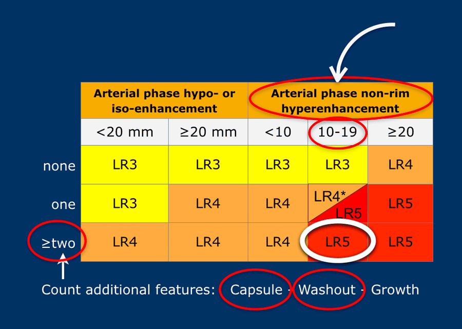
Li Rads Chart - If some, but not all, features of an hcc or benign observation are present then the categories lr 4 (probably hcc) and lr 2 (probably benign) and can be used, respectively. Include capsule in measurement in phase/sequence which margins are clearest. The middle category, lr 3, is used for. Web american college of radiology It includes an updated diagnostic algorithm, a new treatment response assessment algorithm, basic management guidance, basic reporting guidance, key definitions, core supporting material, and faq. It helps eliminate mistakes and improve communication between members of your care team. Do not measure in arterial phase or dwi if margins are clearly visible on different phase. • a comprehensive system for standardizing the acquisition, interpretation, reporting, and data collection of liver imaging Web patient on the list for liver transplant. Cancer which has been biopsied or surgically removed. The term observation is used when it is not sure, whether something is a lesion or a pseudolesion. Do not measure in arterial phase or dwi if margins are clearly visible on different phase. Patient who has received a liver transplant. This method of categorizing liver findings for patients with risk factors for developing hcc allows the radiology community to:. Cancer which has been biopsied or surgically removed. Web the liver imaging reporting and data system (li‐rads) is a comprehensive system that uses standardized terminology, technique, interpretation, and reporting of liver imaging. Benign lesion that did not develop from liver cells and was biopsied or surgically removed. Patient who has received a liver transplant. Do not measure in arterial phase. Web american college of radiology Do not measure in arterial phase or dwi if margins are clearly visible on different phase. Web the liver imaging reporting and data system (li‐rads) is a comprehensive system that uses standardized terminology, technique, interpretation, and reporting of liver imaging. This method of categorizing liver findings for patients with risk factors for developing hcc allows. It includes an updated diagnostic algorithm, a new treatment response assessment algorithm, basic management guidance, basic reporting guidance, key definitions, core supporting material, and faq. If some, but not all, features of an hcc or benign observation are present then the categories lr 4 (probably hcc) and lr 2 (probably benign) and can be used, respectively. The term observation is. It helps eliminate mistakes and improve communication between members of your care team. This method of categorizing liver findings for patients with risk factors for developing hcc allows the radiology community to: Patient who has received a liver transplant. The term observation is used when it is not sure, whether something is a lesion or a pseudolesion. Web patient on. Patient who has received a liver transplant. Web american college of radiology Reduce imaging interpretation variability and errors. The middle category, lr 3, is used for. Web the liver imaging reporting and data system (li‐rads) is a comprehensive system that uses standardized terminology, technique, interpretation, and reporting of liver imaging. The middle category, lr 3, is used for. Web the liver imaging reporting and data system (li‐rads) is a comprehensive system that uses standardized terminology, technique, interpretation, and reporting of liver imaging. If some, but not all, features of an hcc or benign observation are present then the categories lr 4 (probably hcc) and lr 2 (probably benign) and can. Patient who has received a liver transplant. It helps eliminate mistakes and improve communication between members of your care team. It includes an updated diagnostic algorithm, a new treatment response assessment algorithm, basic management guidance, basic reporting guidance, key definitions, core supporting material, and faq. Reduce imaging interpretation variability and errors. The middle category, lr 3, is used for. Web patient on the list for liver transplant. If some, but not all, features of an hcc or benign observation are present then the categories lr 4 (probably hcc) and lr 2 (probably benign) and can be used, respectively. This method of categorizing liver findings for patients with risk factors for developing hcc allows the radiology community to: It includes. Do not measure in arterial phase or dwi if margins are clearly visible on different phase. Reduce imaging interpretation variability and errors. Benign lesion that did not develop from liver cells and was biopsied or surgically removed. It includes an updated diagnostic algorithm, a new treatment response assessment algorithm, basic management guidance, basic reporting guidance, key definitions, core supporting material,. It includes an updated diagnostic algorithm, a new treatment response assessment algorithm, basic management guidance, basic reporting guidance, key definitions, core supporting material, and faq. Web american college of radiology Reduce imaging interpretation variability and errors. Web the liver imaging reporting and data system (li‐rads) is a comprehensive system that uses standardized terminology, technique, interpretation, and reporting of liver imaging. The middle category, lr 3, is used for. Web patient on the list for liver transplant. Benign lesion that did not develop from liver cells and was biopsied or surgically removed. Patient who has received a liver transplant. • a comprehensive system for standardizing the acquisition, interpretation, reporting, and data collection of liver imaging It helps eliminate mistakes and improve communication between members of your care team. If some, but not all, features of an hcc or benign observation are present then the categories lr 4 (probably hcc) and lr 2 (probably benign) and can be used, respectively. The term observation is used when it is not sure, whether something is a lesion or a pseudolesion.
LIRADS Version 2018 Ancillary Features at MRI RadioGraphics

Figure 2 from Cirrhotic liver What's that nodule? The LIRADS approach
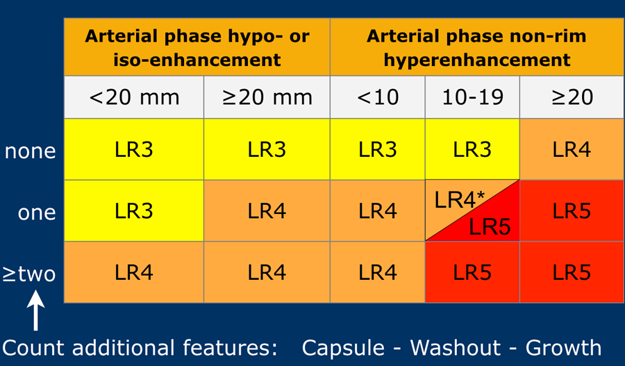
The Radiology Assistant LIRADS
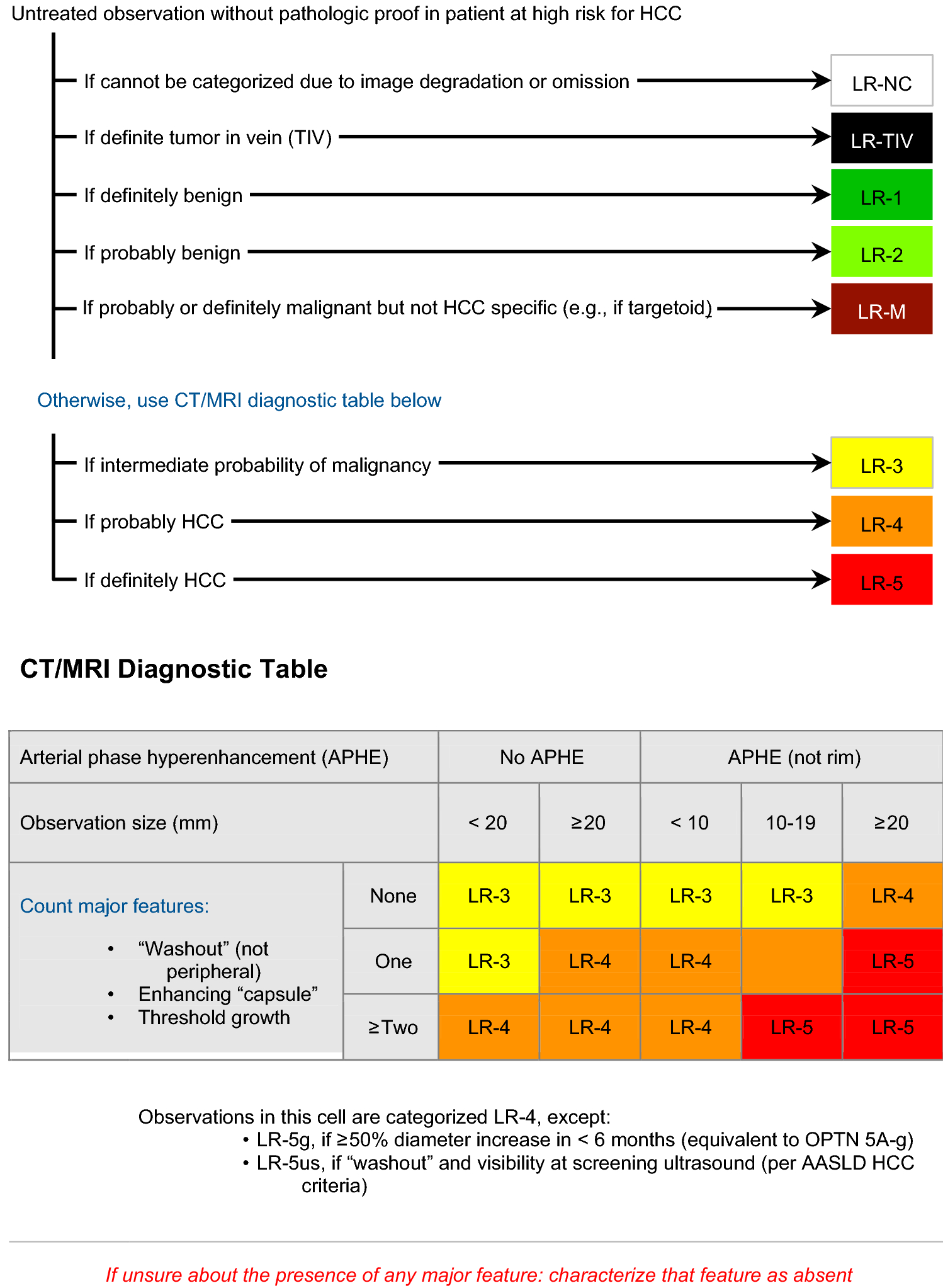
LIRADS version 2018 What is new and what does this mean to my
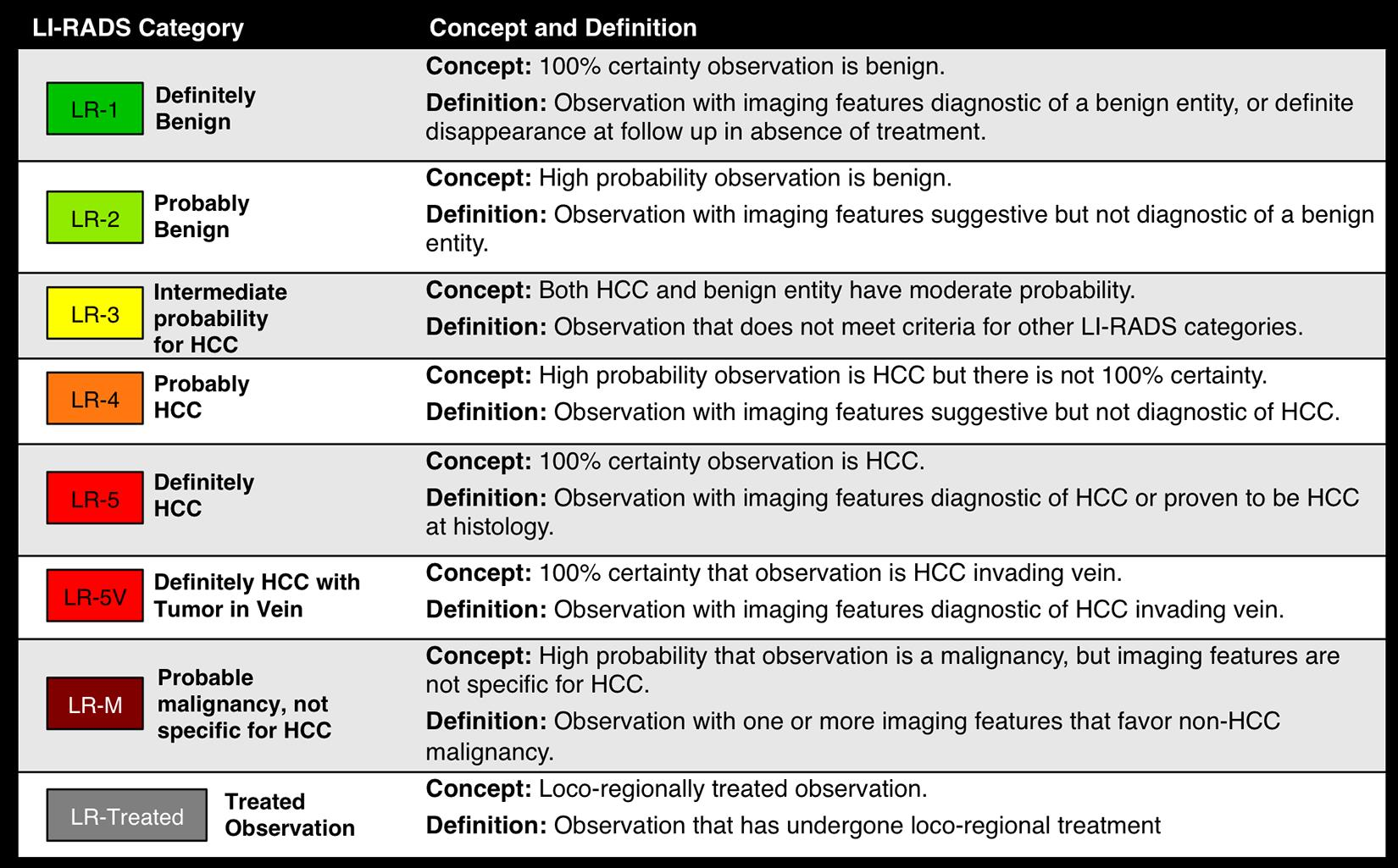
LIRADS Abdominal Imaging Resources

Li Rads Chart
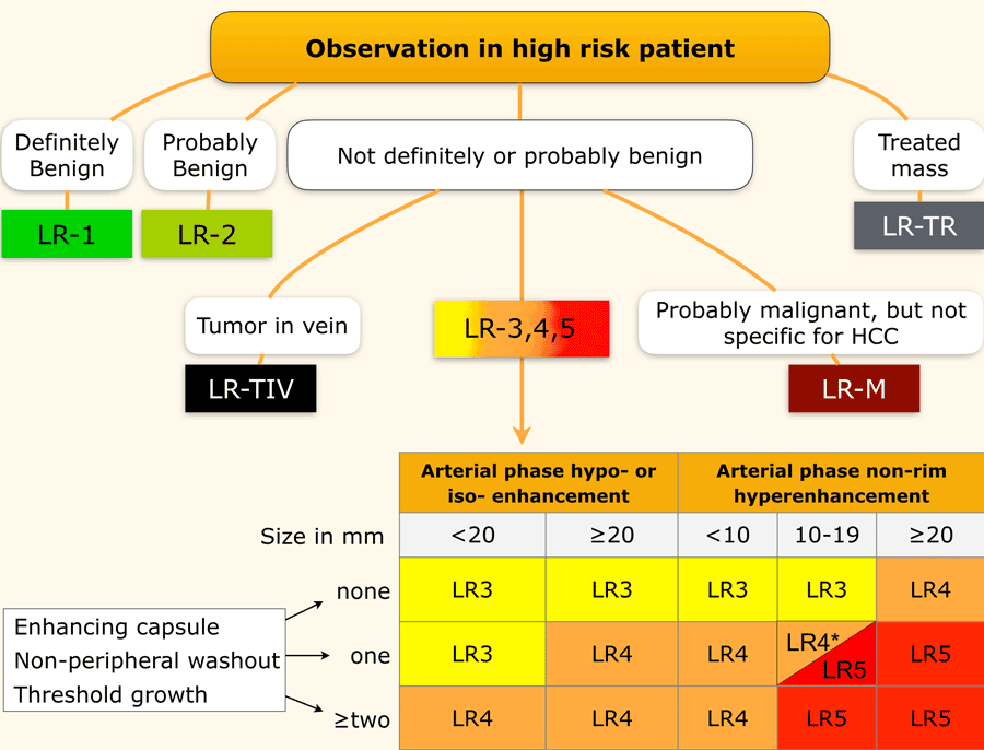
The Radiology Assistant LIRADS
Li Rads Chart

LIRADS for MR Imaging Diagnosis of Hepatocellular Carcinoma

2017 Version of LIRADS for CT and MR Imaging An Update RadioGraphics
Include Capsule In Measurement In Phase/Sequence Which Margins Are Clearest.
Do Not Measure In Arterial Phase Or Dwi If Margins Are Clearly Visible On Different Phase.
Cancer Which Has Been Biopsied Or Surgically Removed.
This Method Of Categorizing Liver Findings For Patients With Risk Factors For Developing Hcc Allows The Radiology Community To:
Related Post: