Labelled Heart Drawing
Labelled Heart Drawing - Web discussed in this video is how to draw and label the structures of the heart, the layers of the heart, and a discussion on how blood flows within the heart. This shape represents the aorta. Right, left, superior, and inferior: Web in this lecture, dr mike shows the two best ways to draw and label the heart! Web muscle and tissue make up this powerhouse organ. The right margin is the small section of the right atrium that extends between the superior and inferior vena cava. The diagram of heart is beneficial for class 10 and 12 and is frequently asked in the examinations. Electrical impulses make your heart beat, moving blood through these chambers. Drag and drop the text labels onto the boxes next to the diagram. Myocardium is the thick middle layer of muscle that allows your heart chambers to contract and relax to pump blood to your body.; Blood transports oxygen and nutrients to the body. This interactive atlas of human heart anatomy is based on medical illustrations and cadaver photography. Your heart contains four muscular sections ( chambers) that briefly hold blood before moving it. The diagram of heart is beneficial for class 10 and 12 and is frequently asked in the examinations. Web anatomy of the. The innermost layer, the endocardium, lines the interior structures of the heart. Web we would like to show you a description here but the site won’t allow us. Start your sketch at the edge of the superior vena cava, and work the shape down to the top edge of the heart’s body. Blood transports oxygen and nutrients to the body.. A 2022 tax filing was filed last year and has been published online. Web the human heart is primarily comprised of four chambers. Drag and drop the text labels onto the boxes next to the diagram. Anatomical illustrations and structures, 3d model and photographs of dissection. The heart is a muscular organ that pumps blood through the blood vessels of. Relate the structure of the heart to its function as a pump. Identify the tissue layers of the heart. Includes an exercise, review worksheet, quiz, and model drawing of an anterior view (frontal section) of the heart in. Web the epicardium covers the heart, wraps around the roots of the great blood vessels, and adheres the heart wall to a. Web we would like to show you a description here but the site won’t allow us. It is also involved in the removal of. Web the cardiovascular system consists of the heart, blood vessels, and the approximately 5 liters of blood that the blood vessels transport. Web labeled heart diagram showing the heart from anterior unlabeled heart diagrams (free download!). Web the heart is located in the thoracic cavity medial to the lungs and posterior to the sternum. Endocardium is the thin inner lining of the heart chambers and also forms the surface of the valves.; Your heart contains four muscular sections ( chambers) that briefly hold blood before moving it. Cardiovascular system animation for u. Web the $130,000 payment. Web muscle and tissue make up this powerhouse organ. Original file (svg file, nominally 663 × 651 pixels, file size: Web the most common heart attack symptoms or warning signs are chest pain, breathlessness, nausea, sweating etc. 93 kb) render this image in. Compare systemic circulation to pulmonary circulation. Blood transports oxygen and nutrients to the body. So if you remember this general pattern, it will help you recall the order in which blood flows through each side of the heart. Myocardium is the thick middle layer of muscle that allows your heart chambers to contract and relax to pump blood to your body.; Right, left, superior, and inferior:. This thick layer is the muscle that contracts to pump and propel blood. Web the most common heart attack symptoms or warning signs are chest pain, breathlessness, nausea, sweating etc. This shape represents the aorta. Base (posterior), diaphragmatic (inferior), sternocostal (anterior), and left and right pulmonary surfaces. It may be a straight tube, as in spiders and annelid worms, or. Describe the internal and external anatomy of the heart. In fishes the heart is a folded tube, with three or four enlarged areas that. Base (posterior), diaphragmatic (inferior), sternocostal (anterior), and left and right pulmonary surfaces. Right, left, superior, and inferior: Web discussed in this video is how to draw and label the structures of the heart, the layers of. This interactive atlas of human heart anatomy is based on medical illustrations and cadaver photography. The user can show or hide the anatomical labels which provide a useful tool to create illustrations perfectly adapted for teaching. Relate the structure of the heart to its function as a pump. It may be a straight tube, as in spiders and annelid worms, or a somewhat more elaborate structure with one or more receiving chambers (atria) and a main pumping chamber (ventricle), as in mollusks. Web the human heart is primarily comprised of four chambers. Web by the end of this section, you will be able to: Both sides work together to efficiently circulate the blood. In this interactive, you can label parts of the human heart. The heart has five surfaces: Size of this png preview of this svg file: In fishes the heart is a folded tube, with three or four enlarged areas that. Web the heart has three layers. Web labeled heart diagram showing the heart from anterior unlabeled heart diagrams (free download!) worksheet showing unlabelled heart diagrams. It also has several margins: Web diagram of the human heart (cropped).svg. Original file (svg file, nominally 663 × 651 pixels, file size: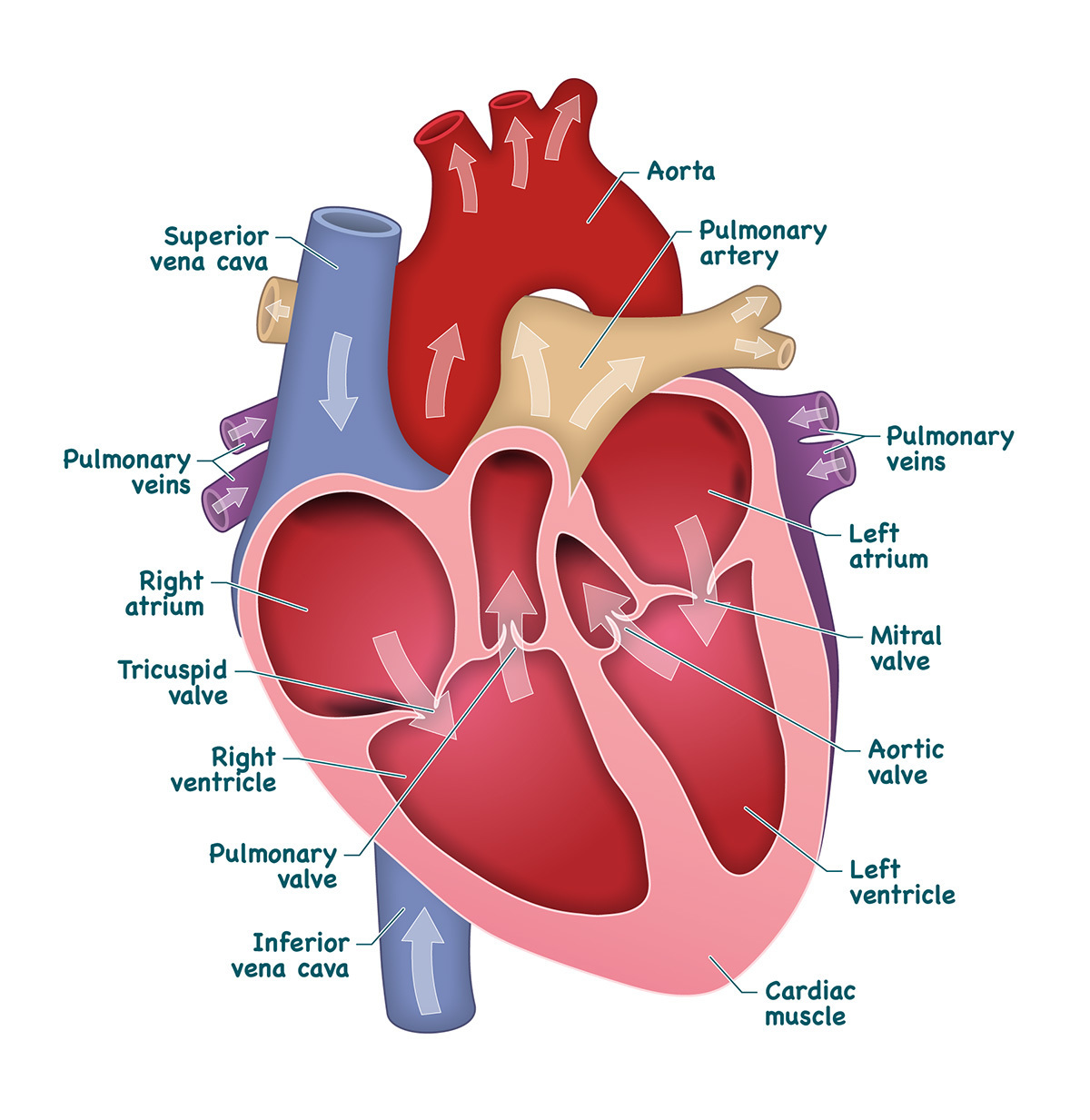
Heart And Labels Drawing at GetDrawings Free download
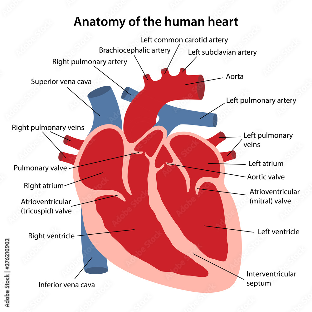
Anatomy of the human heart. Cross sectional diagram of the heart with

Cardiac cycle and the Human Heart A* understanding for iGCSE Biology 2

11+ Heart Drawing With Labels Robhosking Diagram

How to Draw the Internal Structure of the Heart 13 Steps
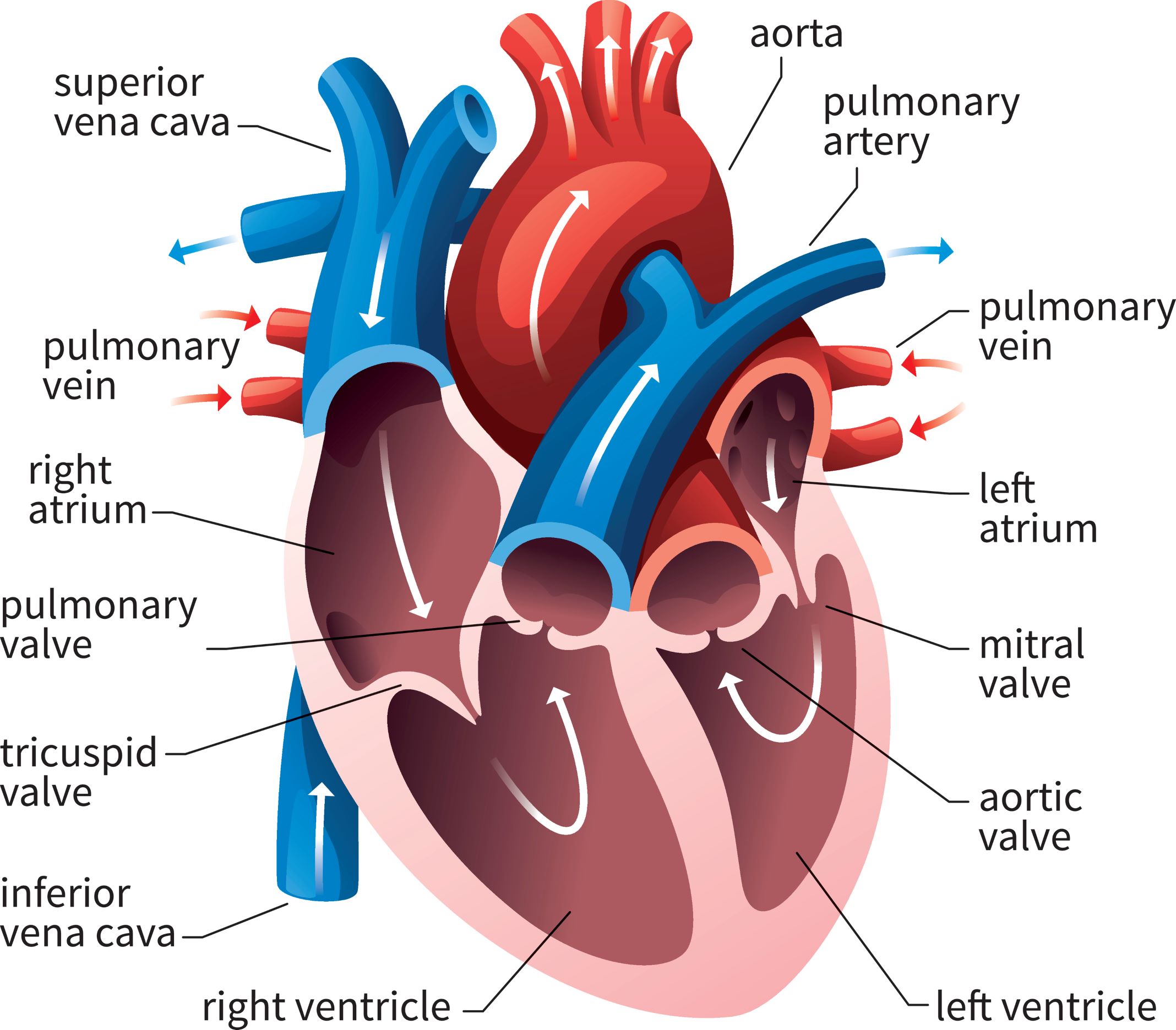
heart anatomy labeling
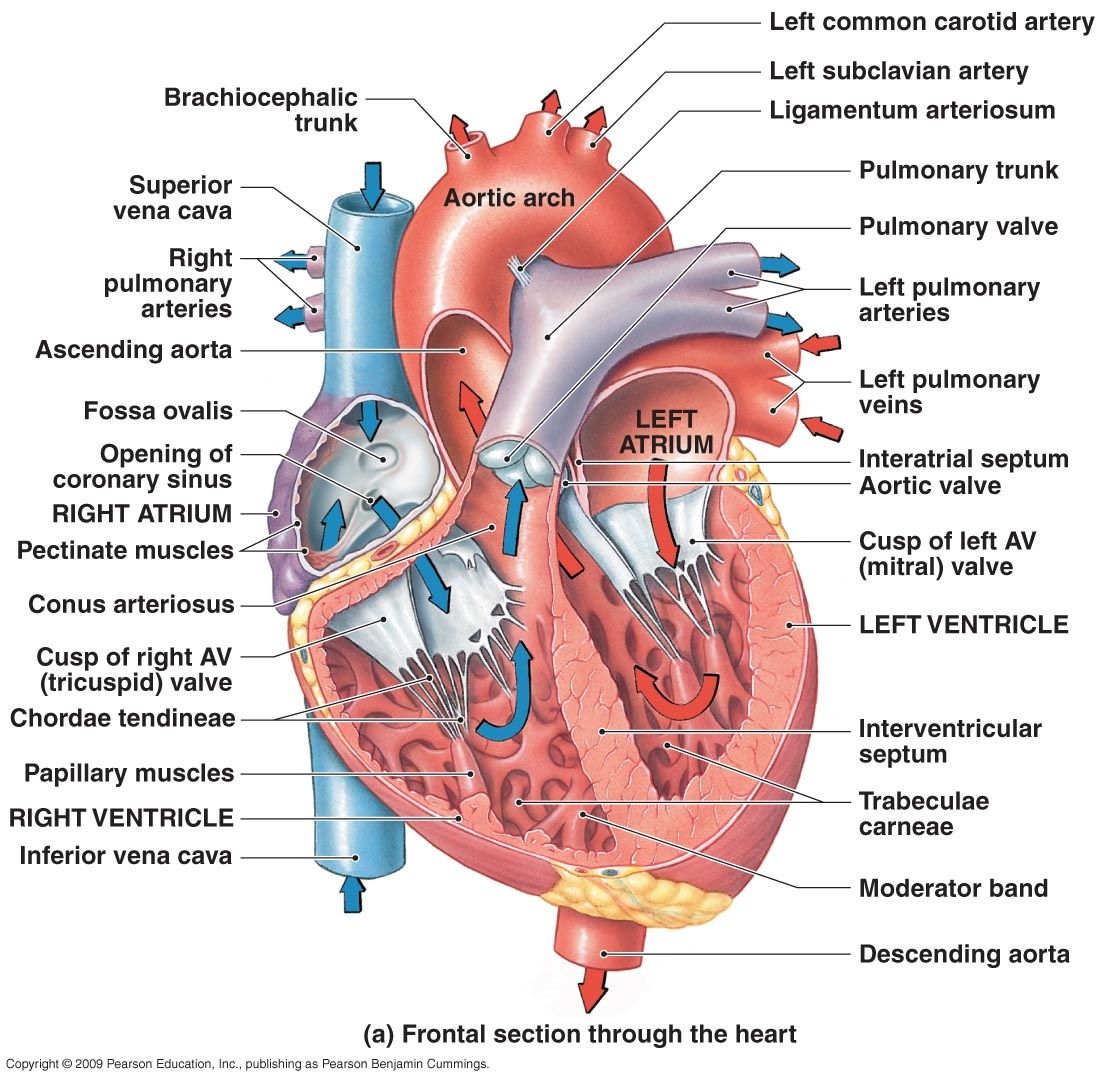
Labeled Drawing Of The Heart at GetDrawings Free download
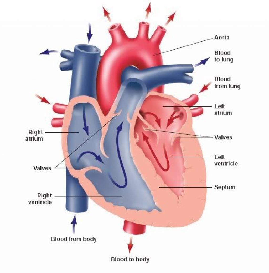
When one teaches, two learn. The heart and the circulatory system

How to Draw the Internal Structure of the Heart 14 Steps
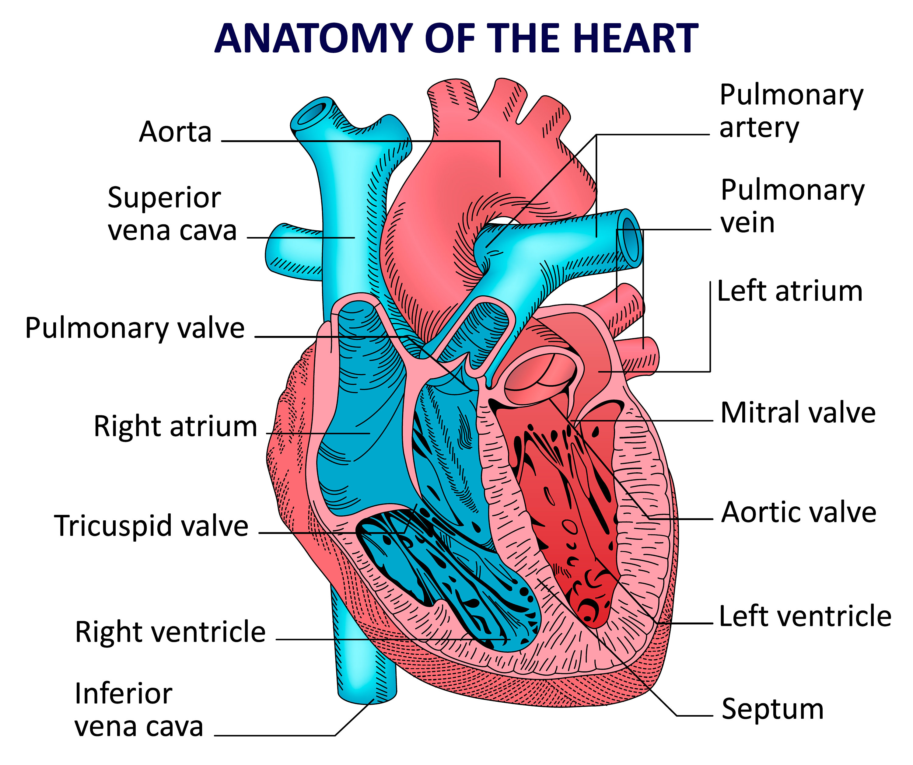
The Human Heart Diagram Labeled
A 2022 Tax Filing Was Filed Last Year And Has Been Published Online.
The Heart Is A Muscular Organ That Pumps Blood Through The Blood Vessels Of The Circulatory System.
Base (Posterior), Diaphragmatic (Inferior), Sternocostal (Anterior), And Left And Right Pulmonary Surfaces.
Web Discussed In This Video Is How To Draw And Label The Structures Of The Heart, The Layers Of The Heart, And A Discussion On How Blood Flows Within The Heart.
Related Post: