Knee Anatomy Drawing
Knee Anatomy Drawing - It helps you stand, move and keep your balance. The femur, tibia and patella. Anterior ligament of fibular head. It allows the lower leg to move relative to the thigh while supporting the body’s weight. It connects your thigh bone (femur) to your shin bone (tibia). Side and front view of knee bones, hand drawn femur, patella, tibia and fibula, tibial plateau and lateral condyle. It is formed by articulations between the patella, femur and tibia. Problems with any part of the knee's anatomy can. Shop best sellersread ratings & reviews Movements at the knee joint are essential to many everyday activities, including walking, running, sitting and standing. Web anatomy of human knee vector sketch of leg bones and joint, medicine design. Web in this video lesson, you will discover how to draw knees from life with the necessary knowledge of this joint's proportions and anatomy. The tibiofemoral joint and patellofemoral joint. This tool is at the same time useful for the training and teaching of the anatomy,. Web the knee joint is a synovial joint that connects three bones; Web the knee joint is one of the strongest and most important joints in the human body. Each anatomical structure was labeled interactively. The knee joint joins the thigh with the leg and consists of two articulations: Anterior intermuscular septum of leg. Anterior ligament of fibular head. The most basic component of knee joint anatomy are the bones which provide the structure to the knee. Movements at the knee joint are essential to many everyday activities, including walking, running, sitting and standing. Shop best sellersread ratings & reviews Anterior superior iliac spine at 1, and anterior inferior iliac spine at. 60 day money backpremium canvas artmade in usa It connects your thigh bone (femur) to your shin bone (tibia). Each anatomical structure was labeled interactively. Anterior ligament of fibular head. One between the femur and tibia, and one between the femur and patella. Shop best sellersread ratings & reviews A pencil drawing of the muscles and the skeleton of a human leg. Web in this episode of simplified constructive anatomy, we cover the structure of the legs and knees. It allows the lower leg to move relative to the thigh while supporting the body’s weight. Content is updated weekly and includes: Web knee anatomy involves more than just muscles and bones. The largest joint in the body, the knee is also one of the most easily injured. Side and front view of knee bones, hand drawn femur, patella, tibia and fibula, tibial plateau and lateral condyle. Anterior superior iliac spine at 1, and anterior inferior iliac spine at. There are four. The same geometry can be applied. Then draw the quadriceps muscles, and indicate the patella and its tendon down to the lower leg. The femur, tibia and patella. Web to draw the knee, begin by visualizing the bones and tendons underneath to help with the placement of landmarks. Contents overview function anatomy conditions and. By understanding the anatomy of the knee we can infer about what is happening underneath when we. Content is updated weekly and includes: Discover (and save!) your own pins on pinterest The front of the leg contains three main muscles that run about parallel from the hip down to the knee. Problems with any part of the knee's anatomy can. It helps you stand, move and keep your balance. The front of the leg contains three main muscles that run about parallel from the hip down to the knee. When the leg is stretched out, the knee joint is placed on a straight line with the hip and ankle (left). Your knees also contain cartilage, like your meniscus, and ligaments,. The front of the leg contains three main muscles that run about parallel from the hip down to the knee. The same geometry can be applied. It is a complex hinge joint composed of two articulations; It helps you stand, move and keep your balance. The knee is a complex joint that flexes, extends, and twists slightly from side to. This tool is at the same time useful for the training and teaching of the anatomy, but also for. The knee is the meeting point of the femur (thigh bone) in the upper leg and the tibia (shinbone) in. Inside larynx nasal throttle anatomy. Then draw the quadriceps muscles, and indicate the patella and its tendon down to the lower leg. Web the knee joint is a hinge type synovial joint, which mainly allows for flexion and extension (and a small degree of medial and lateral rotation). Side and front view of knee bones, hand drawn femur, patella, tibia and fibula, tibial plateau and lateral condyle. Shop best sellersread ratings & reviews Each anatomical structure was labeled interactively. Knee anatomy for figurative artists. The knee joint is one of the largest and most complex joints in the body. Web cartoon illustration of the human knee joint anatomy illustration of the human knee joint anatomy knee joint icon gray illustration on white 3d vector of the human respiratory system, lungs, alveoli. Knee joints, 19 century medical illustration. Movements at the knee joint are essential to many everyday activities, including walking, running, sitting and standing. It helps you stand, move and keep your balance. The snapshot icon at the top center will take a snapshot of your scene that can then be saved as a jpg or drawn on with the included pen tools. It is formed by articulations between the patella, femur and tibia.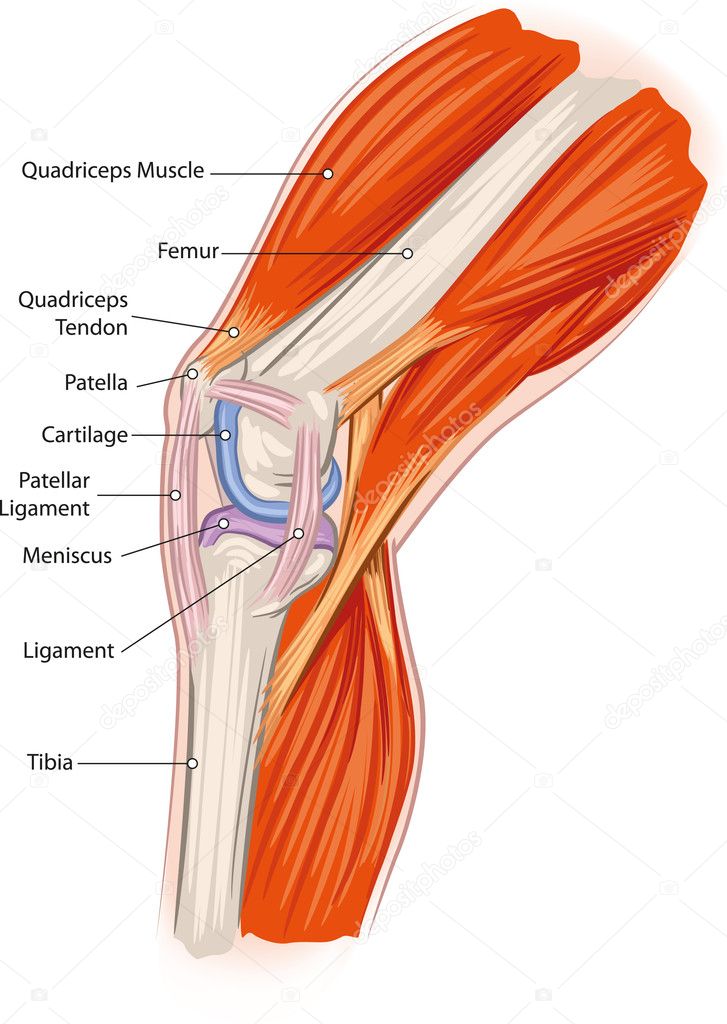
Knee anatomy Stock Vector Image by ©Lukaves 18341225
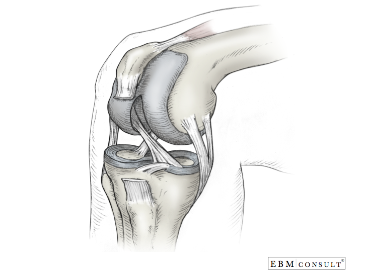
Anatomy Knee
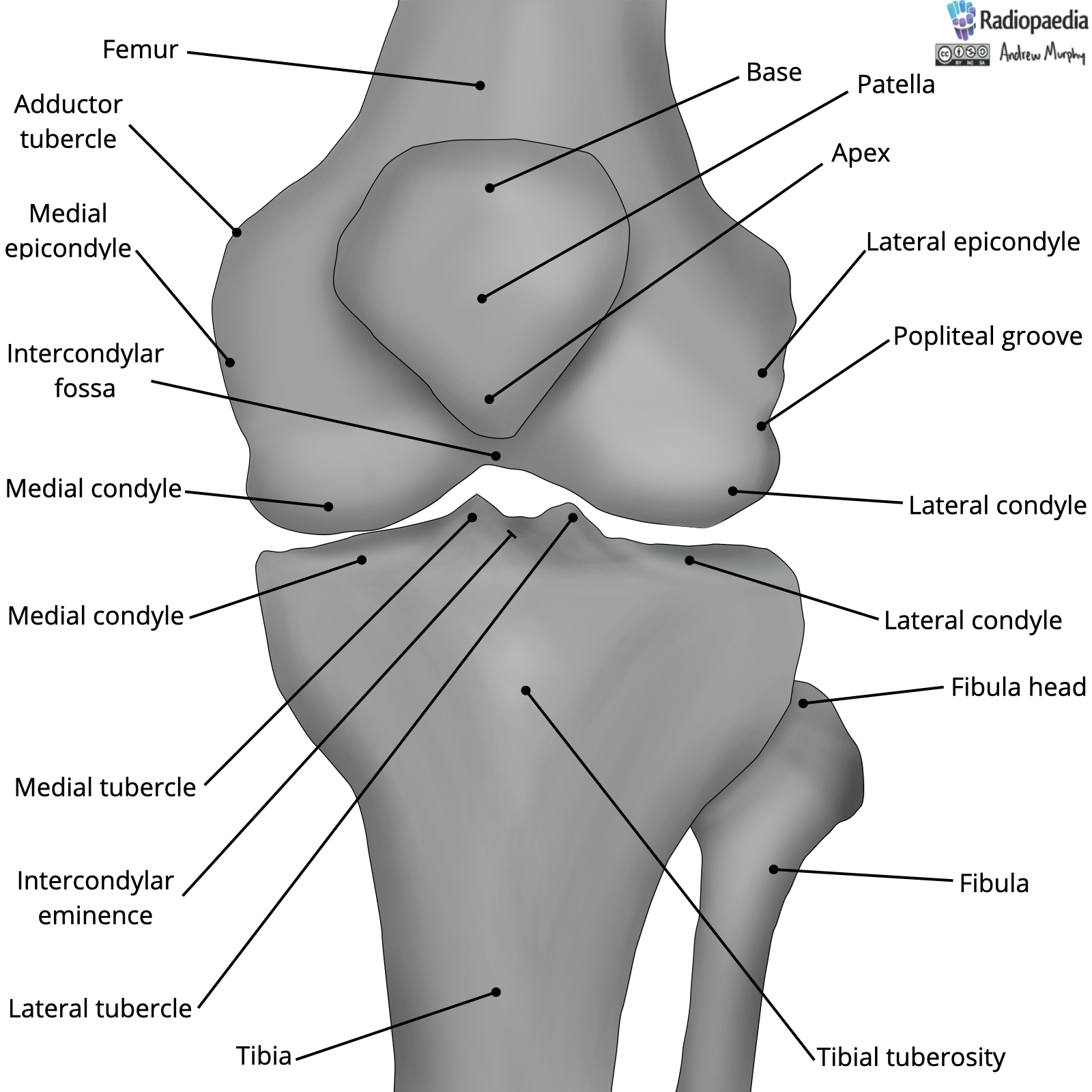
Radiopaedia Drawing Bones of the knee joint English labels

Knee bones and joint sketch human anatomy Vector Image

Structure of the human knee Stock Vector Image by ©Silbervogel 72406683
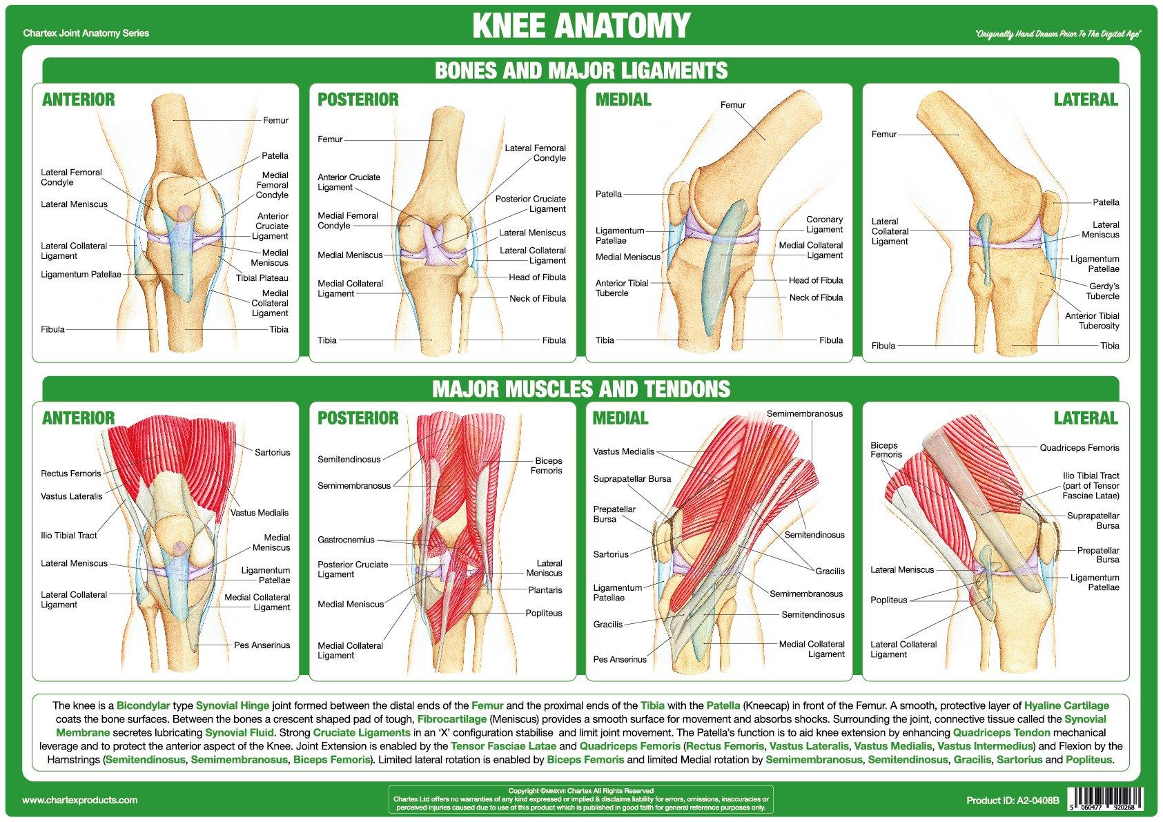
Knee Joint Anatomy Poster

Anatomy of Knee

Knee injuries causes, types, symptoms, knee injuries prevention & treatment
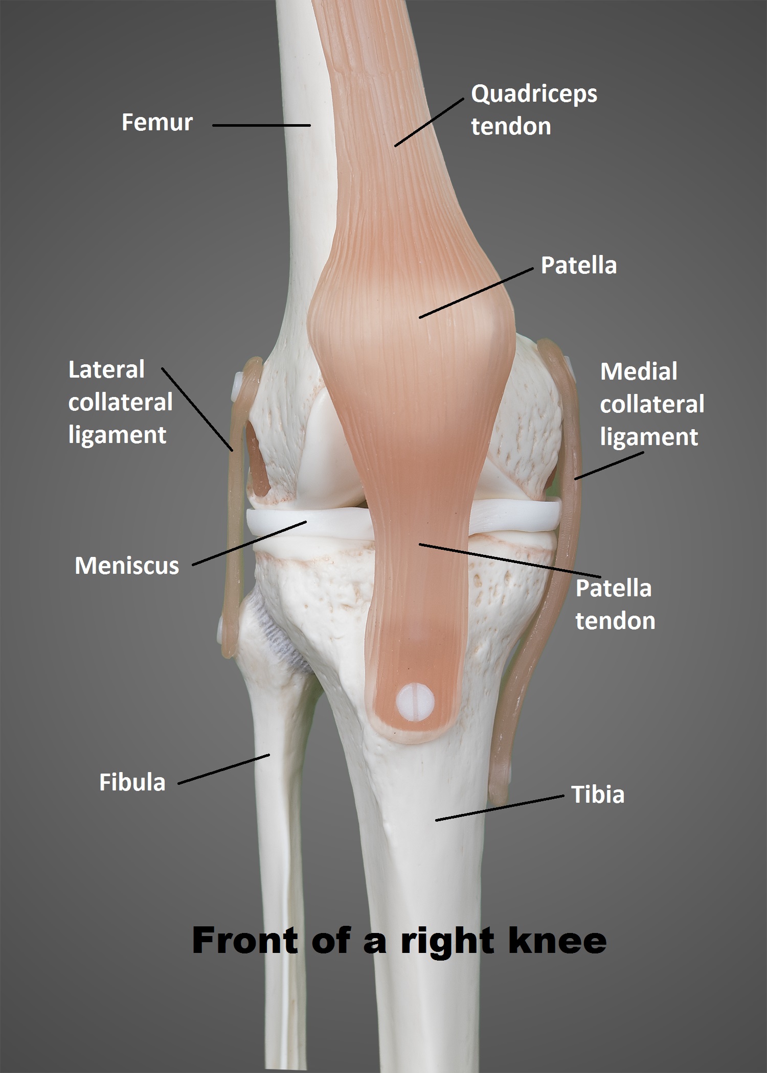
The Knee UT Health San Antonio
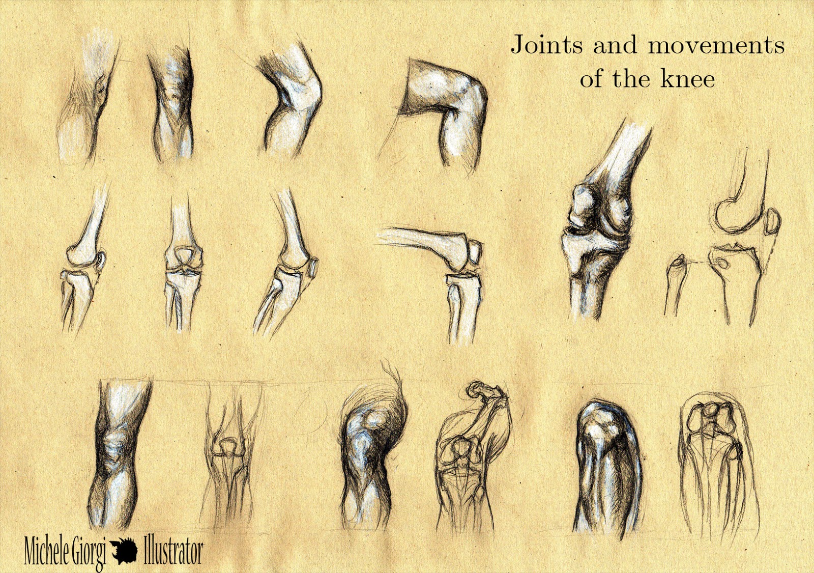
Michele Illustrator Anatomy Sketches Joints and movements of
By Understanding The Anatomy Of The Knee We Can Infer About What Is Happening Underneath When We.
Anatomy Of The Knee For Drawing.
Shop Best Sellersread Ratings & Reviews
It Is A Complex Hinge Joint Composed Of Two Articulations;
Related Post: