Foot And Ankle Anatomy Chart
Foot And Ankle Anatomy Chart - Medial and lateral view of the bones of the foot. The calcaneus is underneath the talus and forms the heel bone. In this article, we shall look at the anatomy of the ankle joint; Identification of compression syndromes requires a very good understanding of the anatomy of the nerves and vessels of the foot and ankle. Web there are a variety of anatomical structures that make up the anatomy of the foot and ankle (figure 1) including bones, joints, ligaments, muscles, tendons, and nerves. Web foot and ankle anatomy consists of 33 bones, 26 joints and over a hundred muscles, ligaments and tendons. Major (2nd most important) medial arch support. Along the bottom, there are three different soles — the two. Covering joint anatomy of the bones, ligaments, muscles, tendons and nerves, plus information on pathologies and common conditions affecting the feet and ankle, our foot anatomical posters are ideal for. Web plantar fascia (windlass mechanism) origin. Web there are a variety of anatomical structures that make up the anatomy of the foot and ankle (figure 1) including bones, joints, ligaments, muscles, tendons, and nerves. Smaller illustrations show the following details: Web the foot and ankle joint consists of 28 bones and is a complex system which provides us with the ability to walk, run and jump.. Web the ankle is the joint that connects your foot to your lower leg. Smaller illustrations show the following details: Illustrates nerve and blood supply to this region, including plantar view of arteries and nerves, common fractures and sprains and anterior impingement syndrome. Web the foot and ankle consists of 26 bones, 33 joints, and many muscles, tendons and ligaments.. Medial and lateral view of the bones of the foot. Web plantar fascia (windlass mechanism) origin. Toward the back of the shoe, you’ll find the: Web there are a variety of anatomical structures that make up the anatomy of the foot and ankle (figure 1) including bones, joints, ligaments, muscles, tendons, and nerves. Web the foot and ankle joint consists. Web foot and ankle anatomy consists of 33 bones, 26 joints and over a hundred muscles, ligaments and tendons. The large central figure shows normal foot and ankle anatomy including bones, muscles and tendons. Web our foot and ankle chart is one of our best selling charts, perfect for learning and explaining the major bony features of the foot and. Web the foot and ankle consists of 26 bones, 33 joints, and many muscles, tendons and ligaments. The distal end of the tibia, the distal end of the fibula, and the superior surface of the talus in the foot. The articulating surfaces, ligaments, movements, and. Along the bottom, there are three different soles — the two. It is made up. These will be reviewed in the sections of this chapter. Covering joint anatomy of the bones, ligaments, muscles, tendons and nerves, plus information on pathologies and common conditions affecting the feet and ankle, our foot anatomical posters are ideal for. This part of the body is able to adapt to uneven terrain, provide shock absorption and allow for even and. This part of the body is able to adapt to uneven terrain, provide shock absorption and allow for even and stable mobility during the gait (walking) cycle. Web foot & ankle charts. Web the foot and ankle joint consists of 28 bones and is a complex system which provides us with the ability to walk, run and jump. Web there. Ebraheim’s educational animated video describes anatomical structures of the foot and ankle, the bony anatomy, the joints, ligaments, and the compartments, in a simple and easy way. Web the foot and ankle consists of 26 bones, 33 joints, and many muscles, tendons and ligaments. In this article, we shall look at the anatomy of the ankle joint; Toward the back. Smaller illustrations show the following details: Learn more about the msd manuals and our commitment to global medical knowledge. Web the ankle joint consists of three bony surfaces: Web our foot and ankle chart is one of our best selling charts, perfect for learning and explaining the major bony features of the foot and ankle. The large central figure shows. The ankle joint connects the leg with the foot, and is composed of three bones: Upper ankle joint (tibiotarsal), talocalcaneonavicular, and subtalar joints. Shows common fractures and sprains and anterior impingement syndrome. The large central figure shows normal foot and ankle anatomy including bones, muscles and tendons. Illustrates nerve and blood supply to this region, including plantar view of arteries. Web our foot and ankle chart is one of our best selling charts, perfect for learning and explaining the major bony features of the foot and ankle. It’s where your shin bone (tibia), calf bone (fibula) and your talus bone meet. Web there are typically about 23 different parts of a shoe. The large central figure shows normal foot and ankle anatomy including bones, muscles and tendons. View our range of foot and ankle anatomy charts and posters, designed by our team of professional medical illustrators. The articulating surfaces, ligaments, movements, and. Toward the back of the shoe, you’ll find the: Smaller illustrations show the following details: Web the ankle is the joint that connects your foot to your lower leg. This complex network of structures fit and work together to bear weight, allow movement and provide a. The last two together are called the lower ankle joint. This ankle poster shows medial and lateral view of the bones and ligaments of the foot and ankle. Upper ankle joint (tibiotarsal), talocalcaneonavicular, and subtalar joints. Within the front half of the shoe, there’s the: Major (2nd most important) medial arch support. Footeducation is committed to helping educate patients about foot and ankle conditions by providing high quality, accurate, and easy to understand information.
This chart shows foot and ankle bone and ligament anatomy, normal
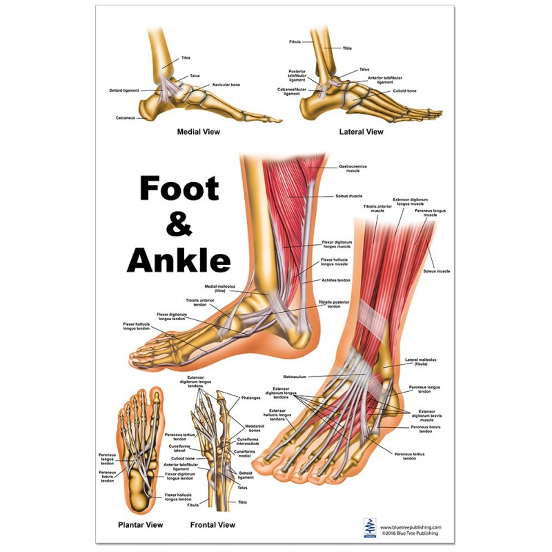
Foot Anatomy Chart
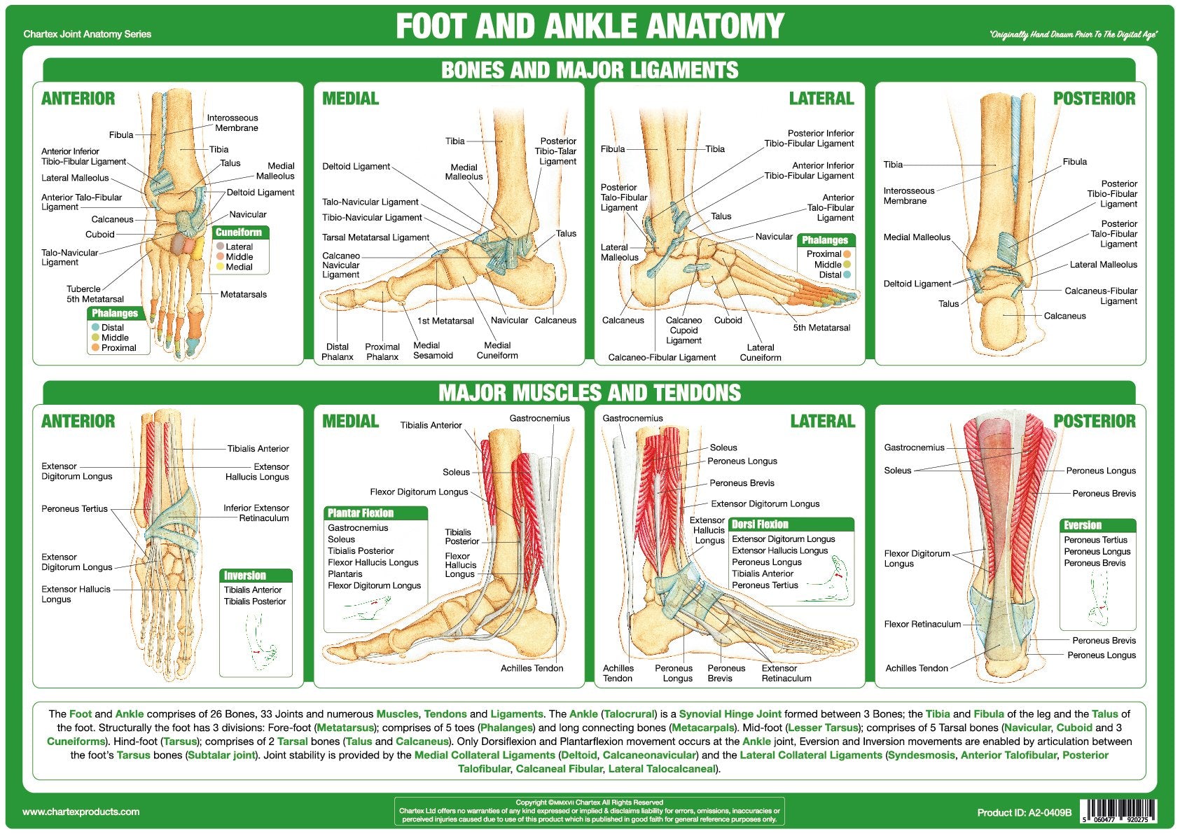
Chartex Foot and Ankle Joint Anatomy Chart

Anatomy of the Foot and Ankle Foot and Ankle Diagram Anatomy of the
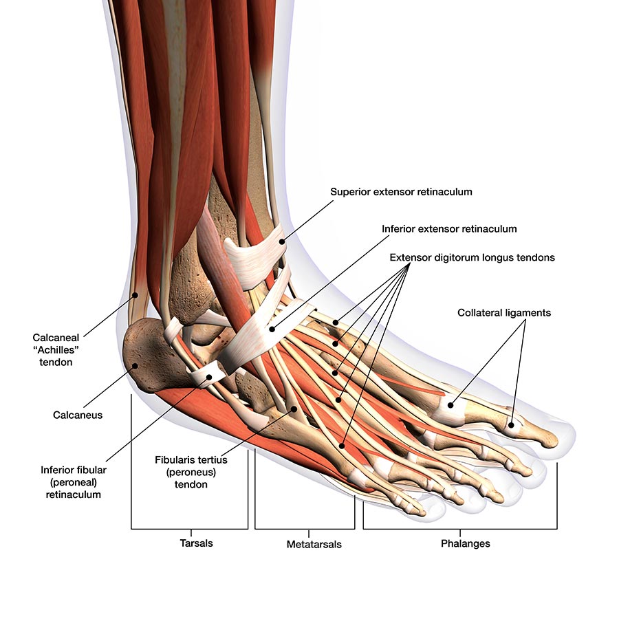
Foot and ankle anatomy, conditions and treatments

Foot & Ankle Anatomy Chart Feet Poster Anatomical Chart
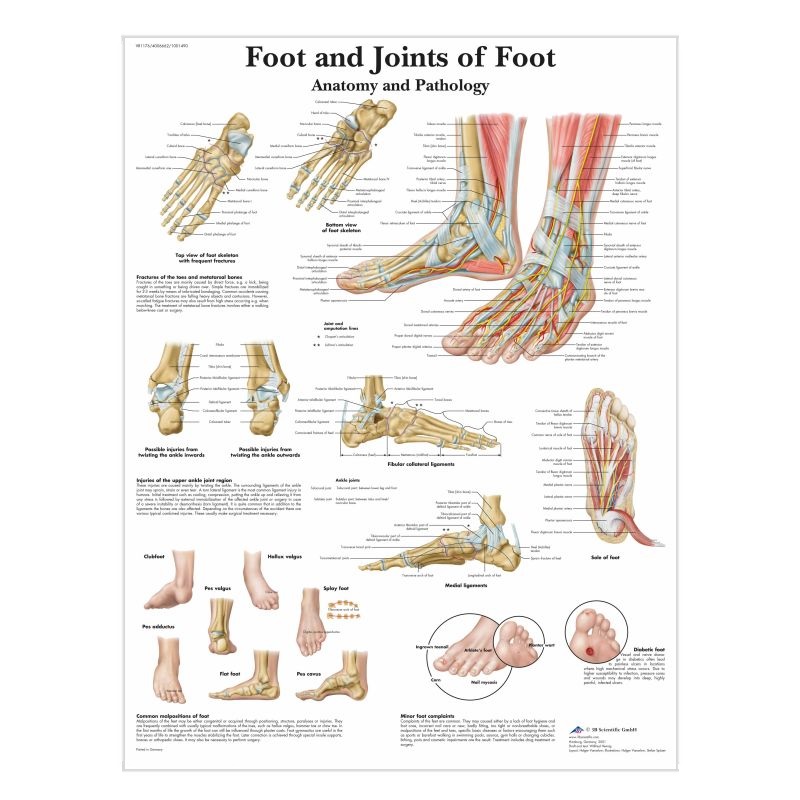
Foot and Ankle Anatomy Chart (Laminated) LabWorld.co.uk
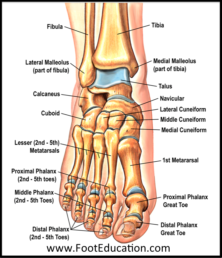
Bones and Joints of the Foot and Ankle Overview FootEducation

Understanding the Foot and Ankle 1004 Anatomical Parts & Charts
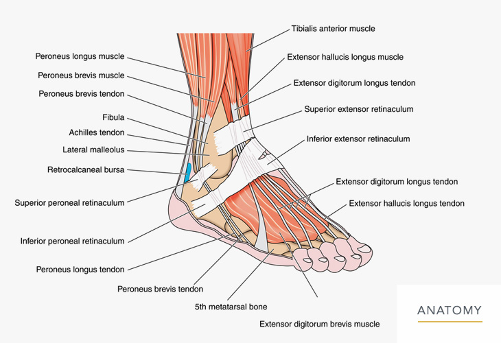
Foot / Ankle Orthopedic Associates of Northern California
The Ankle Joint Connects The Leg With The Foot, And Is Composed Of Three Bones:
The Calcaneus Is Underneath The Talus And Forms The Heel Bone.
Web Toyota Chart Showing Differences In Ankle Anatomy Between Men And Women.
Along The Bottom, There Are Three Different Soles — The Two.
Related Post: