Flow Chart Of A Cell
Flow Chart Of A Cell - Types of user flow charts. Virtually every cell, tissue, organ, and system in the body is impacted by the circulatory system. Flowchart opening statement must be ‘start’ keyword. In a wide range of fields, including software development, engineering, business, and education, it is used to help understand, analyze, and optimize processes. Web a flowchart is a graphical representations of steps. (i) in presence of oxygen. To divide, a cell must complete several important tasks: (ii) in absence of oxygen. Web explain the process of breakdown of glucose in a cell. Flowchart ending statement must be ‘end’ keyword. It must grow, copy its genetic material (dna), and physically split into two daughter cells. Thus the different types of user flows for different purposes. Analysis of a population of cells’ replication state can be achieved by fluorescence labeling of the nuclei of cells in suspension and then analyzing the fluorescence properties of each cell in the population. There are. Preparation of tissue culture cells stored in liquid nitrogen 44. Some textbooks list five, breaking prophase into an early phase (called prophase) and a late phase (called prometaphase). The flow cell has a liquid stream (sheath fluid), which carries and aligns the cells so that they pass single file through the light beam for sensing. But we'll save those specific. Web organelles are involved in many vital cell functions. Every box shows the action taken by the user, while the arrows show the actions taken by the user from one action to the next. This scheme states that information encoded in dna flows into rna via transcription and ultimately to proteins via translation. A flow cell, a measuring system, a. Every box shows the action taken by the user, while the arrows show the actions taken by the user from one action to the next. Virtually every cell, tissue, organ, and system in the body is impacted by the circulatory system. Protocols for preparing cells for flow cytometry 44. A flow cell, a measuring system, a detector, an amplification system,. Preparation of tissue culture cells in suspension 45. Flowchart of a generalized cell culture process. Preparation of adherent tissue culture cell lines 45. The flow cell has a liquid stream (sheath fluid), which carries and aligns the cells so that they pass single file through the light beam for sensing. Web for example, consider the idea that cells are the. Protocols for preparing cells for flow cytometry 44. Web a flowchart is a graphical representation of an algorithm.it should follow some rules while creating a flowchart. It occupies around 95% time of the overall cycle. (ii) in absence of oxygen. If that idea were true, we’d expect to see cells in all kinds of living tissues observed under a microscope. A common task of many research teams is the analysis of cell cycle progression through the distinct cell cycle phases. All symbols in the flowchart must be connected with an arrow line. It was originated from computer science as a tool for representing algorithms and programming logic but had extended to use in all other kinds of processes. Protocols for. Web a flowchart is a type of diagram that visually explains a process or workflow. Protocols for preparing cells for flow cytometry 44. From a cell bank or by isolating cells from donor tissue. This scheme states that information encoded in dna flows into rna via transcription and ultimately to proteins via translation. The flow cell has a liquid stream. Materials and methods we conducted a retrospective cohort study involving patients with stage iib. Nowadays, flowcharts play an extremely important role in displaying information and assisting reasoning. Virtually every cell, tissue, organ, and system in the body is impacted by the circulatory system. Web flow cytometry is a widely used method for analyzing the expression of cell surface and intracellular. Web that is why a user flow is projected in the form of charts with boxes and pointing arrows. Web among these 6,094 cells, 3,839 cells (63%) had at least one rare allele (figure 3k), and this fraction was comparable across the seven blood cell types in the skull (figure 3l). Interphase is the time during which the cell prepares. Web mitosis consists of four basic phases: Web a flowchart is a graphical representations of steps. Web flowcharts are visual representations of processes and systems, showing the various steps, decision points, and paths through a process. If that idea were true, we’d expect to see cells in all kinds of living tissues observed under a microscope — that’s our expected observation. The flow cell has a liquid stream (sheath fluid), which carries and aligns the cells so that they pass single file through the light beam for sensing. Organelles in animal cells include the nucleus, mitochondria, endoplasmic reticulum, golgi apparatus, vesicles, and vacuoles. When starting culture from cells obtained from a cell bank, one needs to go through the procedures of thawing, cell seeding and cell observation. Protocols for preparing cells for flow cytometry 44. Web a data flow diagram (dfd) helps you understand how data flows through a system. Web one of the fundamentals of flow cytometry is the ability to measure the properties of individual particles. By using standardized symbols and definitions, you can create a handy visual representation of any process's various steps and decision points. Flowchart ending statement must be ‘end’ keyword. To divide, a cell must complete several important tasks: Web the basic flow of genetic information in biological systems is often depicted in a scheme known as the central dogma (see figure below). Preparation of tissue culture cells in suspension 45. This scheme states that information encoded in dna flows into rna via transcription and ultimately to proteins via translation.
Parts Of A Cell Flow Chart Kemele

Parts Of A Cell Flow Chart Kemele

What Stimulates Cell Division Brainly Cell cycle presentation by

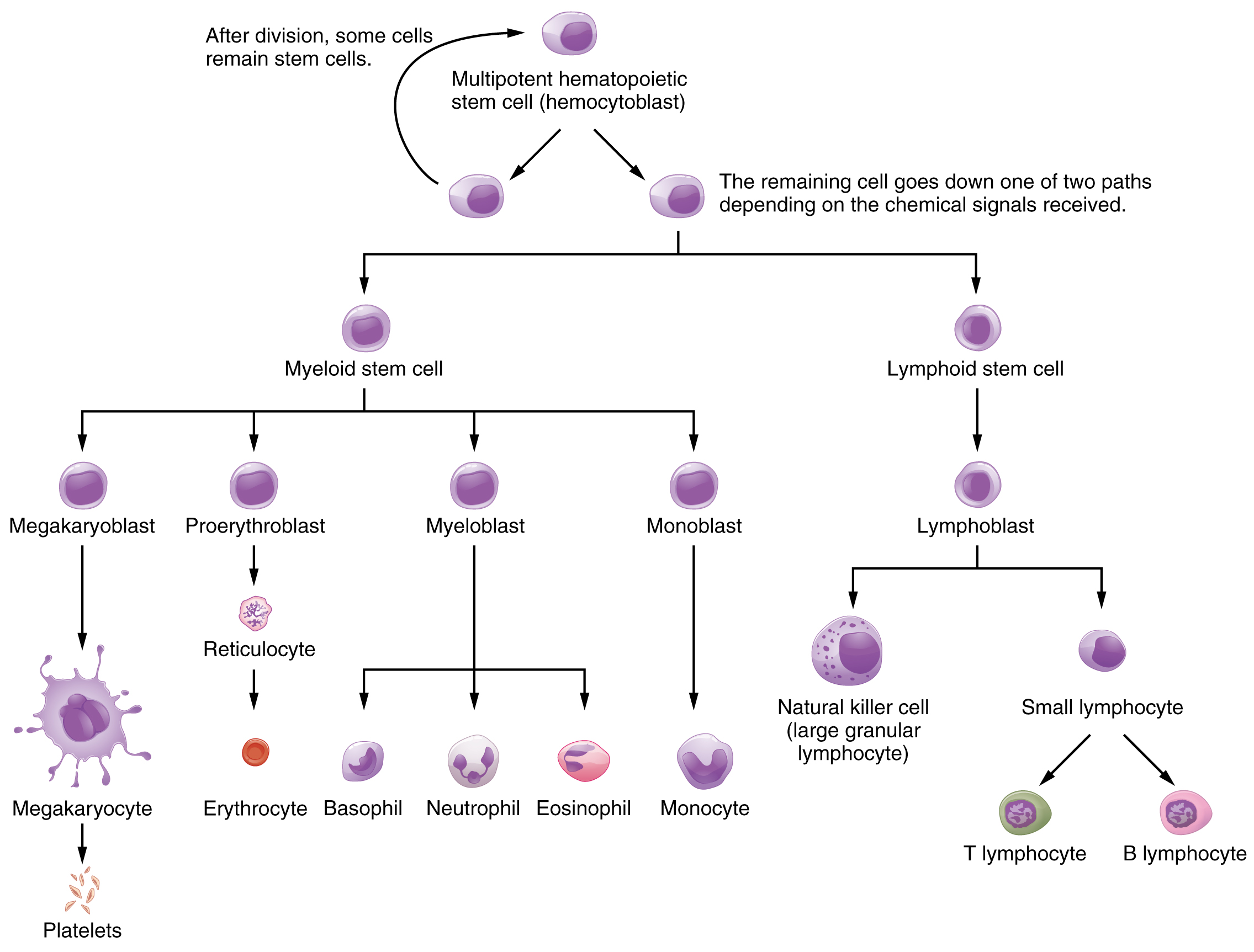
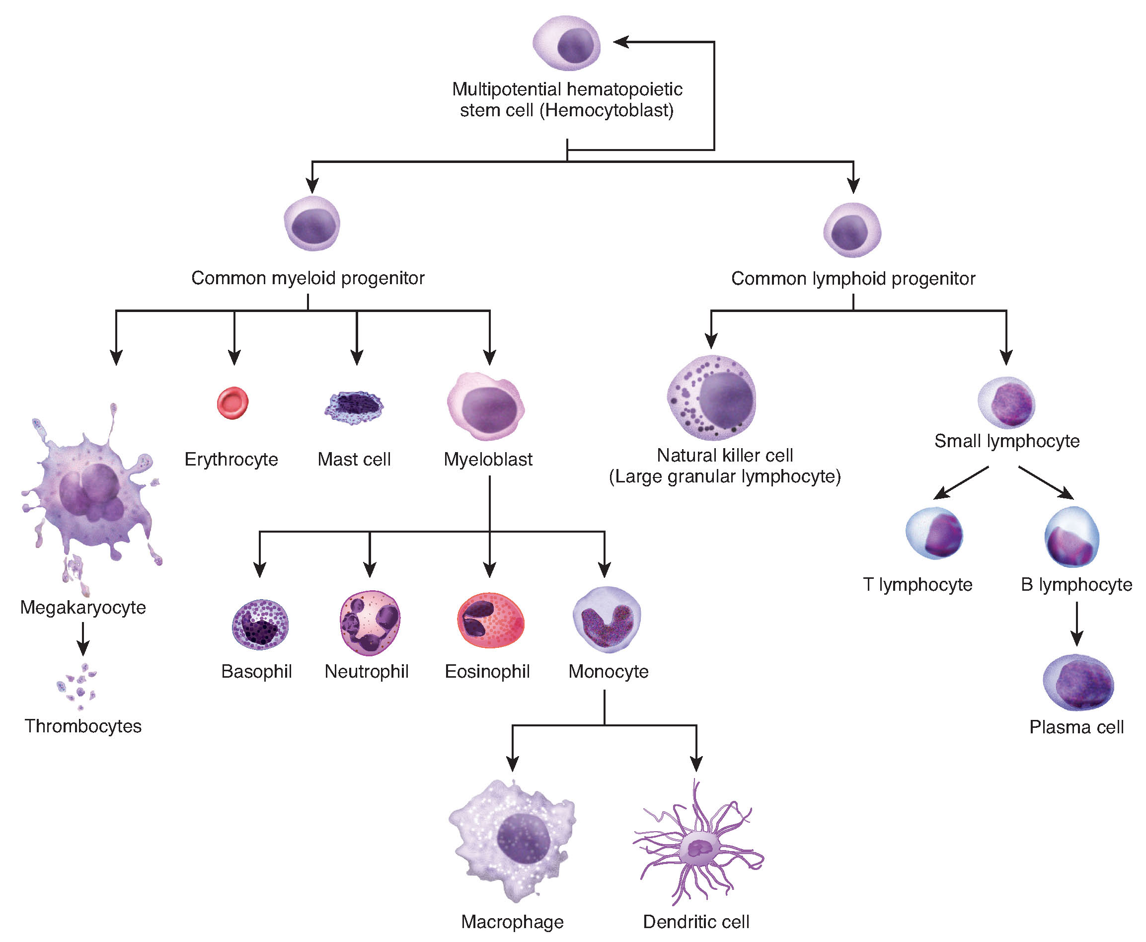
Production of the Formed Elements · Anatomy and Physiology

Cellular Differentiation Anatomy and Physiology I

This flowchart shows the differentiation of a hemocytoblast, a stem

cell and structure flowchart Brainly.in

The flow chart illustrates collection, processing and storage of

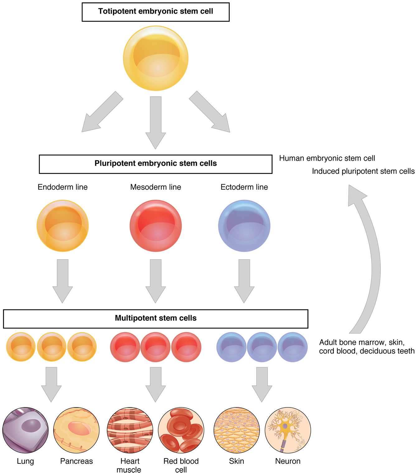
This flow chart shows the differentiation of stem cells into different

Cell Division Flowchart Cell Division
A Dfd Simplifies Complex Processes By Breaking Them Down Into Interconnected Bubbles And Arrows, Creating A Visual Representation Of How Data Moves And Changes.
There Are Two Methods For Obtaining Cells:
Processes Like Reverse Transcription (The Creation Of Dna From And Rna Template.
In A Wide Range Of Fields, Including Software Development, Engineering, Business, And Education, It Is Used To Help Understand, Analyze, And Optimize Processes.
Related Post: