Eye Anatomical Chart
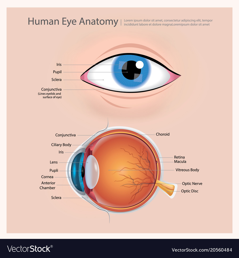
Eye Anatomical Chart - This chart focusses on the anatomy of the eye as it relates to the most frequent causes of eye diseases and is targeted at medical students, trainee ophthalmologists, optometrists, and other eye care specialists. Größe 50,8 x 66 cm. Ein problem mit diesem produkt melden. Anatomy of the extraocular muscles. It contains the fovea, the area of your eye which produces the sharpest images of all. Below, find an explanation of each anatomical part from the above video with physiologic and pathologic correlates. Web orbit (anterior view) the eyes are essential for our daily experience, since about 70% of information we gather is by seeing. The back part of the eye's interior. A clear dome over the iris. Web selecting or hovering over a box will highlight each area in the diagram. Web 6 min read. Mandy helton bas, rvtg, vts(ecc) function of the eye. The pupil is the opening at the center of the iris. This chart focusses on the anatomy of the eye as it relates to the most frequent causes of eye diseases and is targeted at medical students, trainee ophthalmologists, optometrists, and other eye care specialists. Carries the. •acknowledgement of surroundings •communication •help with food acquisition (hunting) •navigation •recognition of friend and foe. It is responsible for producing fine detail in central vision, the part you use when you look directly at something. Each photoreceptor is linked to a nerve fiber. Web what does the macula do? The optic disk, the first part of the optic nerve, is. This chart focusses on the anatomy of the eye as it relates to the most frequent causes of eye diseases and is targeted at medical students, trainee ophthalmologists, optometrists, and other eye care specialists. The back part of the eye's interior. The nerve fibers from the photoreceptors are bundled together to form the optic nerve. Web the optic nerve carries. A diagram to learn about the parts of the eye and what they do. Anatomy of the extraocular muscles. Web the optic nerve carries signals of light, dark, and colors to a part of the brain called the visual cortex, which assembles the signals into images and produces vision. The retina converts light into electrical impulses that are sent to. The optic disk, the first part of the optic nerve, is at the back of the eye. In a number of ways, the. Web human eye, specialized sense organ in humans that is capable of receiving visual images, which are relayed to the brain. Reflexes produced by the streak retinoscope. Here is a tour of the eye starting from the. Web 6 min read. Web the optic nerve carries signals of light, dark, and colors to a part of the brain called the visual cortex, which assembles the signals into images and produces vision. Web overview of the eyes. The opening in the middle of the iris through which light passes to the back of the eye. Anatomy of the. Clear and flexible, the lens changes shape to focus light on the retina. Below, find an explanation of each anatomical part from the above video with physiologic and pathologic correlates. The iris is the colored part of the eye that regulates the amount of light entering the eye. Ein problem mit diesem produkt melden. The chart covers general anatomy of. Interactive ophthalmic figures for medical student education illustrate concepts in eye anatomy and functions in an engaging format. The macula is a small highly sensitive part of the retina. Anatomical eye models and charts for optometrists and patient education. The chart covers general anatomy of the eye with colorful detailed renderings all fully labeled. Here is a tour of the. The opening in the middle of the iris through which light passes to the back of the eye. Web here are descriptions of some of the main parts of the eye: Clear and flexible, the lens changes shape to focus light on the retina. It is responsible for producing fine detail in central vision, the part you use when you. Your eye is a slightly asymmetrical globe, about an inch in diameter. The optic disk, the first part of the optic nerve, is at the back of the eye. The outermost layer, known as the fibrous tunic , is composed of the cornea and sclera , which provide shape to the eye and support the deeper structures. Anatomy & embryology. A clear dome over the iris. The opening in the middle of the iris through which light passes to the back of the eye. Web human eye, specialized sense organ in humans that is capable of receiving visual images, which are relayed to the brain. The pupil is the opening at the center of the iris. Only the most important anatomical details are listed, alongside web links to videos and diagrams (each underlined text is a link) external eye and adnexa. The macula is a small highly sensitive part of the retina. Web eye anatomy (16 parts of the eye & what they do) the following are parts of the human eyes and their functions: Web what does the macula do? The lens is a clear part of the eye behind the iris that helps to The outermost layer, known as the fibrous tunic , is composed of the cornea and sclera , which provide shape to the eye and support the deeper structures. Web 6 min read. Web unique 3d anatomical perspectives of the eye. Anatomy warehouse provides a comprehensive selection of human eye models and charts, each displaying the inner structures of the eye. Your eye is a slightly asymmetrical globe, about an inch in diameter. Web selecting or hovering over a box will highlight each area in the diagram. The cornea is the clear outer part of the eye’s focusing system located at the front of the eye.
Labeled Diagram Of Eye
/GettyImages-1128675065-e4bac15b0f39449dba31f25f1020bc8a.jpg)
Eye Anatomy Poster
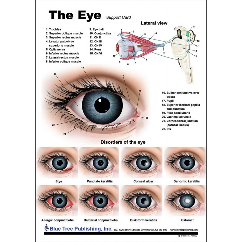
Eye Anatomical Chart

Eye Diagram Vector Art, Icons, and Graphics for Free Download
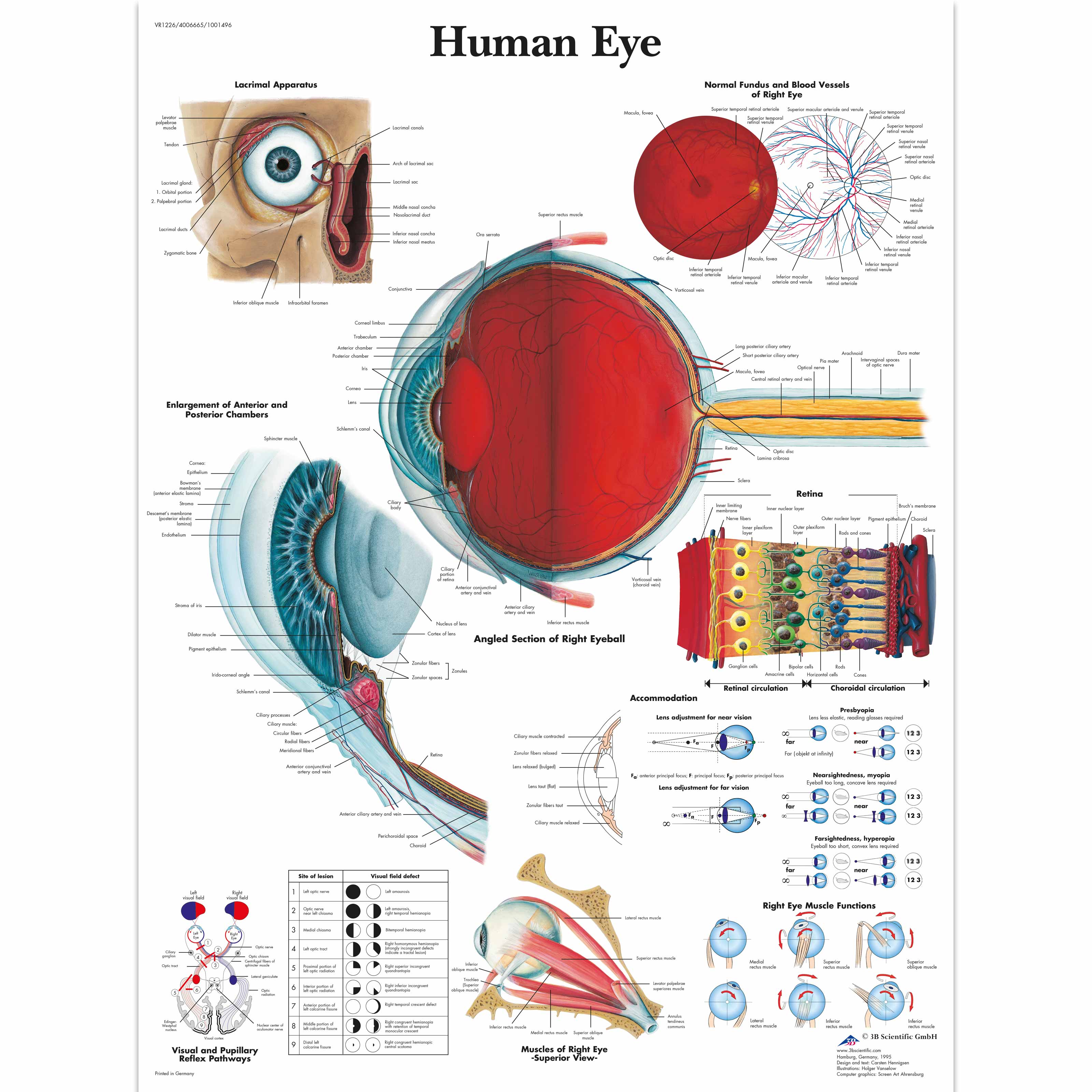
Human Eye Chart 1001496 3B Scientific VR1226L Ophthalmology
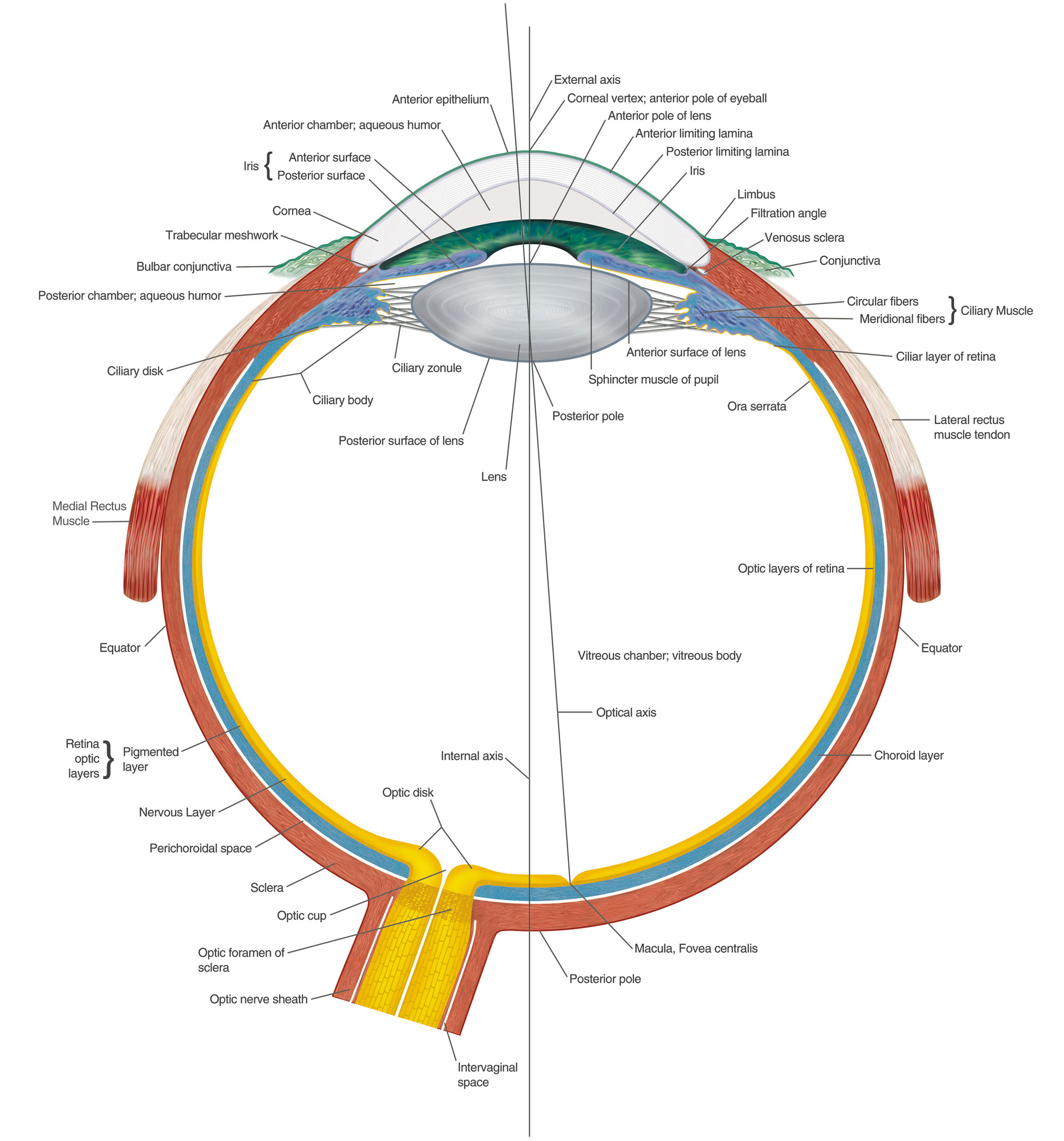
Eye Anatomy Chart B
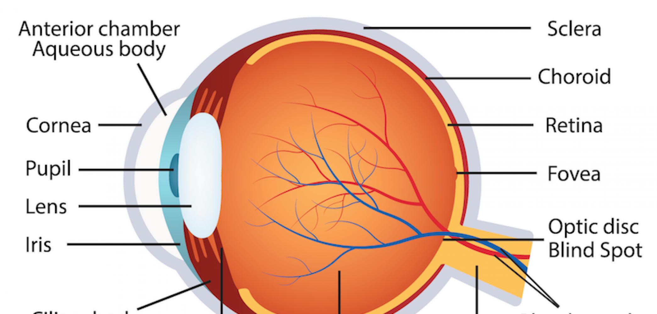
humaneyeanatomy La Pine Eyecare Clinic
Eye Anatomy Poster

Human eye anatomy Royalty Free Vector Image VectorStock

Understanding The Eye Anatomy Health Life Media
Clear And Flexible, The Lens Changes Shape To Focus Light On The Retina.
Größe 50,8 X 66 Cm.
•Acknowledgement Of Surroundings •Communication •Help With Food Acquisition (Hunting) •Navigation •Recognition Of Friend And Foe.
Web To Understand The Diseases And Conditions That Can Affect The Eye, It Helps To Understand Basic Eye Anatomy.
Related Post: