Drawing Of Mitosis
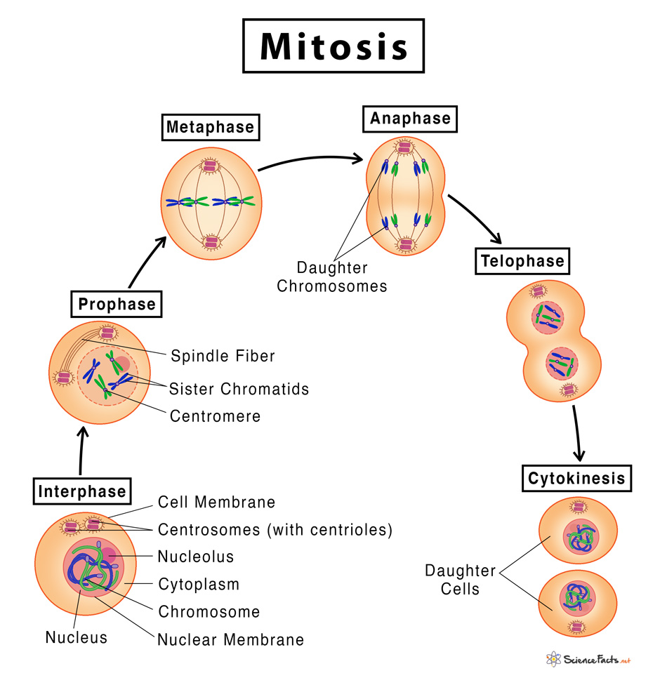
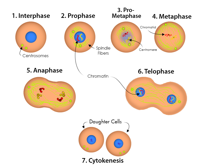
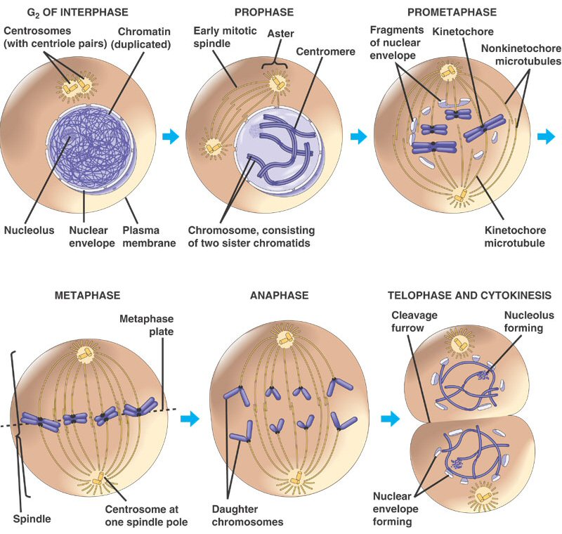
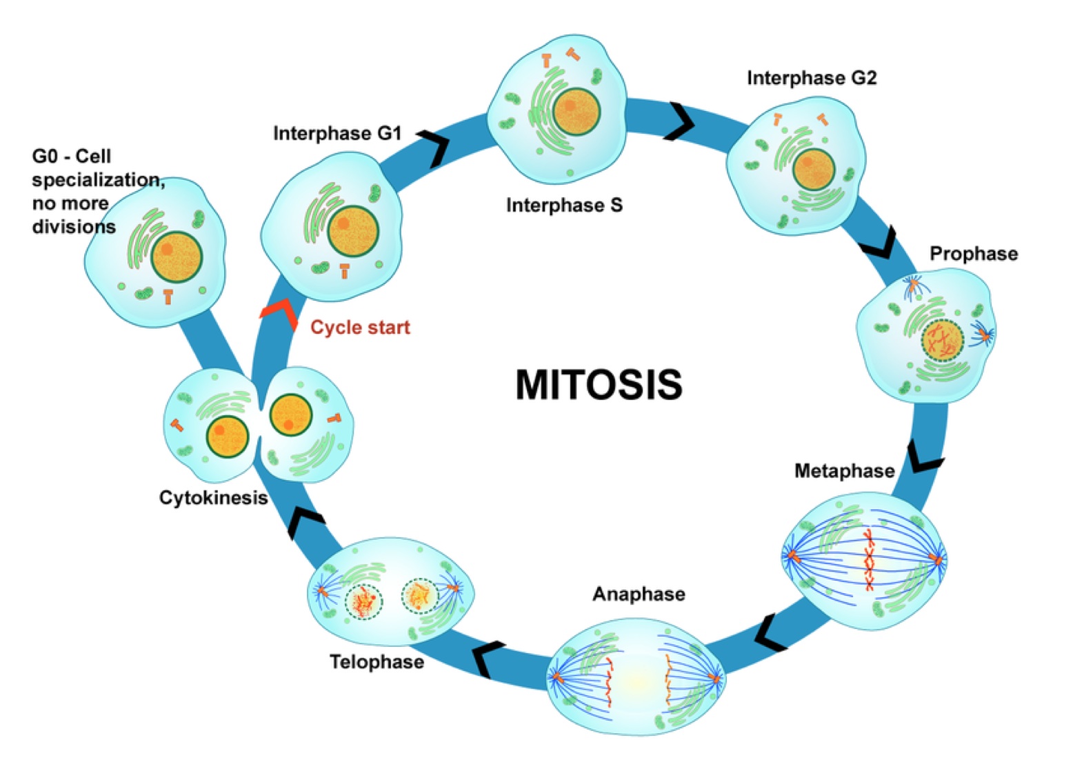
Drawing Of Mitosis - It works by copying each chromosome, and then separating the copies to different sides of the cell. This image is linked to the following scitable pages: Chromatin begins to condense into distinguishable chromosomes. Mitosis, a key part of the cell cycle, involves a series of stages (prophase, metaphase, anaphase, and telophase) that facilitate cell division and genetic information transmission. The micrographs below are onion (allium cepa) root tip cells. The end of prophase is marked by the beginning of the organization of a group of fibres to form a spindle and the disintegration of the nuclear membrane. Web locate the region of active cell division, known as the root apical meristem, which is about 1 mm behind the actual tip of the root. The nuclear membrane is intact. Web how to draw the stages of mitosis and what are the points that you need to take care of while drawing these stages. During mitosis, chromosomes will align, separate, and move into new daughter cells. Figure 1 by openstax college, biology ( cc by 3.0 ). Web today, mitosis is understood to involve five phases, based on the physical state of the chromosomes and spindle. This video also lists the features of eac. When the cell division process is complete, two daughter cells with identical genetic material are produced. | learn science at scitable. Identify and draw a cell in each of the four stages of mitosis in the onion slide. Web today, mitosis is understood to involve five phases, based on the physical state of the chromosomes and spindle. Web phase ends when mitosis begins. The bulk of the cell cycle is spent in the “living phase”, known as. The cell cycle refers. Prophase, metaphase, anaphase, and telophase. The stages of the cell cycle are: Some textbooks list five, breaking prophase into an early phase (called prophase) and a late phase (called prometaphase). Web mitosis diagram showing the different stages of mitosis. Individual chromosomes are not visible. Web below is an illustration and a corresponding micrograph for each stage in mitosis, showing a hypothetical plant cell where 2n=4 (two sets of chromosomes, two chromosomes per set). This is when the cell grows and copies its dna before moving into mitosis. Individual chromosomes are not visible. These phases are prophase , prometaphase , metaphase, anaphase , and. While. That way, when the cell divides down the middle, each new cell gets its own copy of each chromosome. The stages of mitosis are: The nuclear membrane is intact. The g 1 , s, and g 2 phases together are known as interphase. Web mitosis is the process in which a eukaryotic cell nucleus splits in two, followed by division. A cell plate separates the daughter cells. Mitosis in a whitefish embryo. Web locate the region of active cell division, known as the root apical meristem, which is about 1 mm behind the actual tip of the root. Web phase ends when mitosis begins. Identify and draw a cell in each of the four stages of mitosis in the onion. Figure 1 by openstax college, biology ( cc by 3.0 ). Web locate the region of active cell division, known as the root apical meristem, which is about 1 mm behind the actual tip of the root. This image is linked to the following scitable pages: Web in cytokinesis, animal cells: Web mitosis diagram showing the different stages of mitosis. Web in cytokinesis, animal cells: The cell cycle refers to a series of events that describe the metabolic processes of growth and replication of cells. Dna is uncondensed and in the form of chromatin. Mitosis is the process of nuclear division by which two genetically identical daughter nuclei are produced that are also genetically identical to the parent cell nucleus. Dna is uncondensed and in the form of chromatin. The g 1 , s, and g 2 phases together are known as interphase. Web in cytokinesis, animal cells: Web survey the slide to find a cell in each phase of mitosis. The end of prophase is marked by the beginning of the organization of a group of fibres to form. Individual chromosomes are not visible. Web mitosis diagram showing the different stages of mitosis. Interphase is the longest part of the cell cycle. Web survey the slide to find a cell in each phase of mitosis. The stages of mitosis are: This video also lists the features of eac. Web the concept of mitosis. In eukaryotic cells, the cell cycle is divided into two major phases: The nucleolus , a rounded structure, shrinks and disappears. These phases are prophase , prometaphase , metaphase, anaphase , and. It works by copying each chromosome, and then separating the copies to different sides of the cell. Web mitosis consists of four basic phases: Now that we’ve reviewed each of the steps, let’s look at the cycle as a whole: Every base pair of their. Web survey the slide to find a cell in each phase of mitosis. The five phases of mitosis and cell. A cell plate separates the daughter cells. Draw a cell for each phase below. A cleavage furrow separates the daughter cells. During mitosis, chromosomes will align, separate, and move into new daughter cells. Some textbooks list five, breaking prophase into an early phase (called prophase) and a late phase (called prometaphase).
Mitosis Definition, Stages, & Purpose, with Diagram

FileMitosis schematic diagramen.svg Wikimedia Commons

Mitosis stages Diagram drawing CBSE easy way Labeled Science

Mitosis Introduction to Mitosis Mitosis Explained with Diagram
/GettyImages-5304586361-59dfd070845b3400116b5d8b.jpg)
Understand the Stages of Mitosis and Cell Division

Mitosis Diagram by Mulsivaas on DeviantArt

What is mitosis? Facts

Mitosis Royaleb's Blog

Mitosis

6 Stages Mitosis Vector & Photo (Free Trial) Bigstock
Individual Chromosomes Are Not Visible.
The Micrographs Below Are Onion (Allium Cepa) Root Tip Cells.
Web In Cytokinesis, Animal Cells:
The Purpose Of Mitosis Is To Make More Diploid Cells.
Related Post: