Dog Ear Cytology Chart
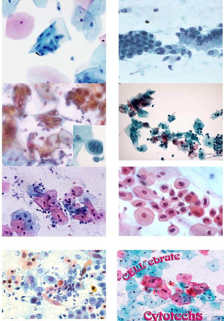
Dog Ear Cytology Chart - Cytology is an important diagnostic technique allowing assessment of the presence of bacteria and yeast on the skin surface and in the external ear canals. Collecting, processing, and evaluating ear cytology samples are skills all credentialed veterinary technicians and nurses learn in credentialing programs.1 ear. Identify clinical lesions that would indicate skin cytology should be performed. Use these images to review common findings present on ear cytology. Web the canine ear consists of the pinna, external ear canal, middle ear and inner ear. Web ear cytology is a test that can help clinicians diagnose and treat otitis externa. When to perform ear cytology, how to collect and stain samples, and some of the common findings veterinary nurses encounter in dogs and cats. 3 i prefer the slide to be heat fixed, then stained with a quick stain (eg, diffquik). Web rods or mixed infection with rods and cocci are identified on cytology. The swab is then rolled onto a microscopic slide until the specimen is evenly distributed ( figure 1 ). Identification of an underlying cause of infection is vital, especially where the otitis is recurrent or chronic. Identify clinical lesions that would indicate skin cytology should be performed. The auricular cartilage of the pinna becomes funnel shaped at the opening of the external ear canal. Web ear cytology is one of the most important investigative steps in all cases of. The auricular cartilage of the pinna becomes funnel shaped at the opening of the external ear canal. Also, in cases of mixed infection, cytology assists in evaluating the relative significance of each organism, strengthening interpretation of culture and sensitivity data. Then work your way up to a high magnification, remembering to use oil immersion for the xloo lens, which is. Use these images to review common findings present on ear cytology. When to perform ear cytology, how to collect and stain samples, and some of the common findings veterinary nurses encounter in dogs and cats. Cytology is an important diagnostic technique allowing assessment of the presence of bacteria and yeast on the skin surface and in the external ear canals.. Samples for cytologic examination are obtained by gently inserting cotton swabs into the horizontal ear canal. Web ear cytology can be simple, rapid and inexpensive, and samples are relatively easy to obtain from conscious dogs. “ear cytology is a quick and easy diagnosis and management tool,” mr williams adds. Web at this stage a cytology test can be performed to. It is used for diagnostic and for treatment success monitoring purposes. Web otitis externa is a common presentation in small animal veterinary practice and the veterinary nurse can play a vital role in managing these cases. The auricular cartilage of the pinna becomes funnel shaped at the opening of the external ear canal. Cytology is an important diagnostic technique allowing. The aim of these articles is to provide veterinary nurses with a basic overview of some cytological sampling and preparation techniques and provide a few pictures of common cells seen in cytology. Stained and unstained samples are useful to assess the ear for the presence respectively of bacterial and yeast pathogens and ectoparasites. Otitis extema is an acute or chronic. Cytology samples from the external ear canal are evaluated for the presence of mites and the presence and numbers of yeast, bacteria, and leukocytes. Web place under microscope and starting with a low magnification, find a densely purple area as this is where inflammatory cells are more likely to be. Primary disorders initiate the inflammatory process within the ear canal. Samples for cytologic examination are obtained by gently inserting cotton swabs into the horizontal ear canal. Stained and unstained samples are useful to assess the ear for the presence respectively of bacterial and yeast pathogens and ectoparasites. Web the canine ear consists of the pinna, external ear canal, middle ear and inner ear. Web ear cytology is a noninvasive, effective. Stained and unstained samples are useful to assess the ear for the presence respectively of bacterial and yeast pathogens and ectoparasites. Infection has failed to respond to appropriate antibiotics. Also, in cases of mixed infection, cytology assists in evaluating the relative significance of each organism, strengthening interpretation of culture and sensitivity data. Web collect cytology from the skin and ears. Identify clinical lesions that would indicate skin cytology should be performed. Web the canine ear consists of the pinna, external ear canal, middle ear and inner ear. Identification of an underlying cause of infection is vital, especially where the otitis is recurrent or chronic. Web at this stage a cytology test can be performed to indicate infection type. Web this. Cytology samples from the external ear canal are evaluated for the presence of mites and the presence and numbers of yeast, bacteria, and leukocytes. Web cytology can characterize the severity of overgrowth or infection; The swab is then rolled onto a microscopic slide until the specimen is evenly distributed ( figure 1 ). Samples for cytologic examination are obtained by gently inserting cotton swabs into the horizontal ear canal. • sample the less severely afected ear first. 3 i prefer the slide to be heat fixed, then stained with a quick stain (eg, diffquik). Infection has failed to respond to appropriate antibiotics. Before staining check under 4x for ear mites. Identification of primary causes and predisposing and perpetuating factors. Collecting, processing, and evaluating ear cytology samples are skills all credentialed veterinary technicians and nurses learn in credentialing programs.1 ear. Web ear cytology is a test that can help clinicians diagnose and treat otitis externa. The external ear is composed of auricular and annular cartilage. Otitis extema is an acute or chronic inflammation of the external ear canal. Sampling, processing, and microscopic evaluation. Stained and unstained samples are useful to assess the ear for the presence respectively of bacterial and yeast pathogens and ectoparasites. Web ear cytology is one of the most important investigative steps in all cases of otitis externa and lends itself well to input from the veterinary nurse.
Animal Ear Cytology Identification Chart

Ear Cytology Dog Chart

Recurrent Ear Infections (Otitis Externa) The Skin Vet

Dog Ear Cytology Rods

Animal Ear Cytology Identification Chart
Veterinary Dog Ear Cytology Chart

Crash Course in Cytology 101 So...I've had several flicker… Flickr

Canine Ear Cytology Chart

Canine Ear Cytology Chart
Dog ear infection yeast vs. bacterial
Web The Canine Ear Consists Of The Pinna, External Ear Canal, Middle Ear And Inner Ear.
Identify Clinical Lesions That Would Indicate Skin Cytology Should Be Performed.
Primary Disorders Initiate The Inflammatory Process Within The Ear Canal And Alter The Aural Environment, Allowing Secondary Complicating Factors, Such As Infections, To Develop.
Griffin Et Al Found That Heat Fixing Versus Not Heat Fixing Otic Exudate On Glass Slides Before Staining Did Not Increase Or Decrease The Number Of Malassezia Yeast Organisms Found On Cytologic Evaluation.
Related Post:
