Diaphragm Drawing
Diaphragm Drawing - Web is the diaphragm an organ. Graphic illustration of lungs organ visualization. It is attached anteriorly to the xiphoid process and costal margin, laterally to the 11th and 12th ribs, and posteriorly to the lumbar vertebrae.the posterior attachment to the vertebrae is by tendinous bands called the. Web satisfyer curvy 3+. Web to practice, inhale through your nose, allow your belly and diaphragm (the muscle at the bottom of your ribs) to expand, exhale through your nose, and draw your belly in. The diaphragm is located between the thoracic and abdominal cavities [3], with important organs like the. Request price add to basket remove add to board. Web the diaphragm is a muscle that helps you breathe. Web glass cosmetic flask with four compartments (kohl tube) of 1. Web the diaphragm is a muscular partition that separates the thoracic cavity from the abdominal cavity in the human body. All rights to paintings and other images found on paintingvalley.com are owned by their respective owners (authors, artists), and the. Graphic illustration of lungs organ visualization. Web is the diaphragm an organ. The diaphragm is a musculotendinous structure with a peripheral attachment to a number of bony structures. Air within the lungs is forced out of the body as the. It is an important structure that plays a crucial role in respiration, as it is responsible for generating the negative pressure that helps to draw air into the lungs. 47.6 mb (4.0 mb compressed) 3334 x 4987 pixels. Please contact your account manager if you have any query. Web learning diaphragmatic breathing. Turquoise template palette, copy space for text. Turquoise template palette, copy space for text. Please contact your account manager if you have any query. Graphic illustration of lungs organ visualization. 28.2 x 42.2 cm · 11.1 x 16.6 in (300dpi) this image is not available for purchase in your country. Graphic illustration of lungs organ visualization. Air within the lungs is forced out of the body as the size of the thoracic cavity decreases. Web 43+ free diaphragm illustrations. Please contact your account manager if you have any query. 47.6 mb (4.0 mb compressed) 3334 x 4987 pixels. Graphic illustration of lungs organ visualization. Human internal organ lungs disease research and recovery. Designed to simulate the sucking or tapping sensation of oral sex, clitoral suction toys are a relatively recent addition to the. Breathing exercises can strengthen your diaphragm and keep it working like. To complete the image in three dimensions, we are left to our own devices. Diaphragm drawing stock photos are available. Web the thoracic cavity becomes deeper and larger, drawing in air from the atmosphere. Web origin and insertion. Web to practice, inhale through your nose, allow your belly and diaphragm (the muscle at the bottom of your ribs) to expand, exhale through your nose, and draw your belly in. Designed to simulate the sucking or tapping sensation of oral sex,. Web satisfyer curvy 3+. Human internal organ lungs disease research and recovery. It serves two main functions: Web the diaphragm is a thin skeletal muscle that sits at the base of the chest and separates the abdomen from the chest. This creates more space in your chest cavity, allowing the lungs. Graphic illustration of lungs organ visualization. It is attached anteriorly to the xiphoid process and costal margin, laterally to the 11th and 12th ribs, and posteriorly to the lumbar vertebrae.the posterior attachment to the vertebrae is by tendinous bands called the. Request price add to basket remove add to board. Many conditions, injuries and diseases can affect how the diaphragm. Choose from 144 diaphragm drawing stock illustrations from istock. Air within the lungs is forced out of the body as the size of the thoracic cavity decreases. Human internal organ lungs disease research and recovery. It is an important structure that plays a crucial role in respiration, as it is responsible for generating the negative pressure that helps to draw. 47.6 mb (4.0 mb compressed) 3334 x 4987 pixels. The human chest cavity, opened from the front, with the lungs and the heart (without a pericardium). It is attached anteriorly to the xiphoid process and costal margin, laterally to the 11th and 12th ribs, and posteriorly to the lumbar vertebrae.the posterior attachment to the vertebrae is by tendinous bands called. Many conditions, injuries and diseases can affect how the diaphragm works, causing symptoms such as trouble breathing and chest pain. It is a skeletal muscle [3], and like many other important muscles, the diaphragm is an organ, one of the most important respiratory organs that humans cannot live without [4]. Web cute young yogi girl with twelve hands in lotus position does belly vacuum and muscle wave, vector illustration with black ink contour lines isolated on a white background in cartoon and hand drawn style. Web browse 190+ diaphragm drawing stock photos and images available, or start a new search to explore more stock photos and images. Turquoise template palette, copy space for text. Web origin and insertion. It contracts and flattens when you inhale. Air within the lungs is forced out of the body as the size of the thoracic cavity decreases. It is an important structure that plays a crucial role in respiration, as it is responsible for generating the negative pressure that helps to draw air into the lungs. All rights to paintings and other images found on paintingvalley.com are owned by their respective owners (authors, artists), and the. When you inhale, your diaphragm contracts (tightens) and moves downward. Where is the diaphragm located. Graphic illustration of lungs organ visualization. Web the diaphragm is a thin skeletal muscle that sits at the base of the chest and separates the abdomen from the chest. Human internal organ lungs disease research and recovery. Web learning diaphragmatic breathing.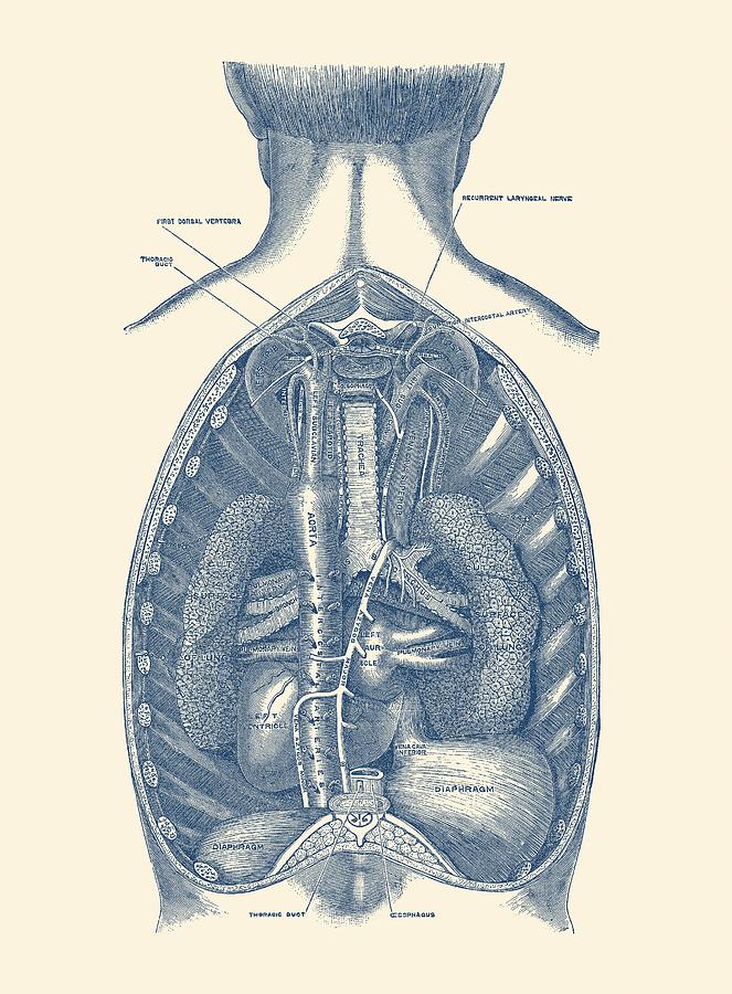
Diaphragm Human Anatomy Rear View Drawing by Vintage Anatomy Prints
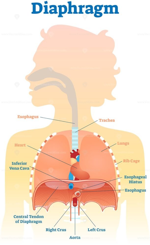
Diaphragm anatomical vector illustration diagram VectorMine
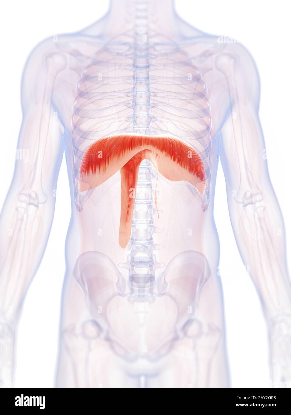
3d rendered illustration of the human diaphragm Stock Photo Alamy
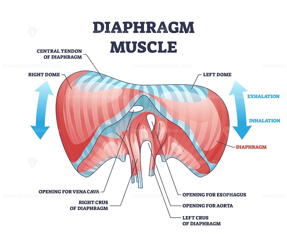
Diaphragm muscle with exhalation and inhalation movement outline

Diaphragm, Drawing Stock Image C024/4326 Science Photo Library
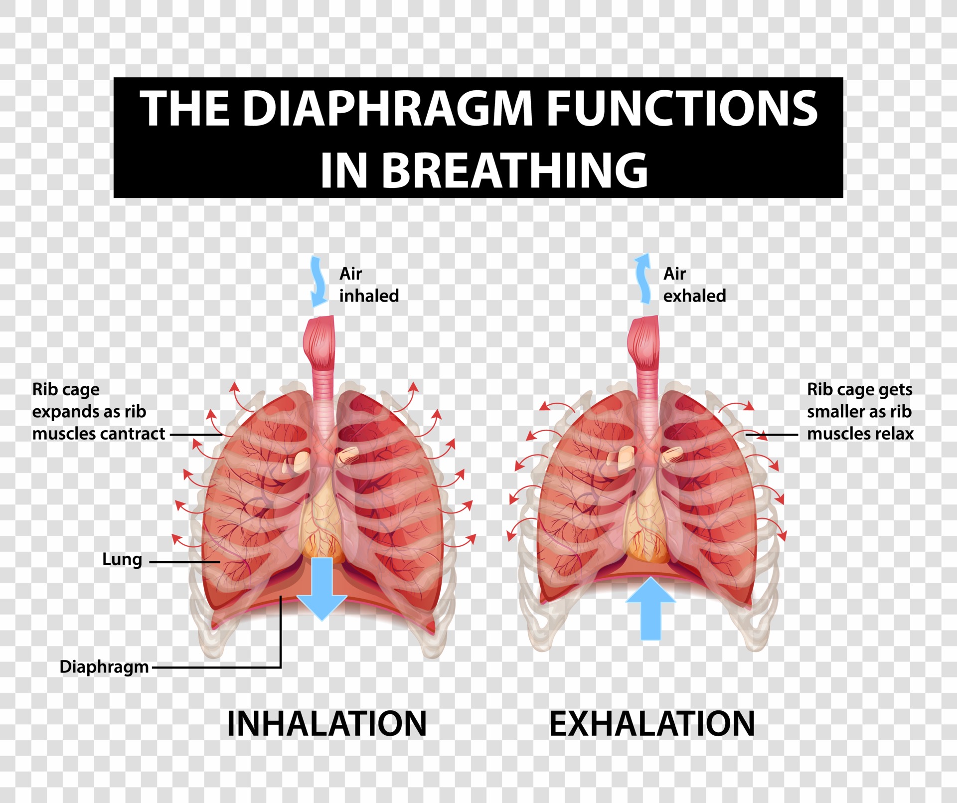
Diagram showing diaphragm functions in breathing 2747522 Vector Art at
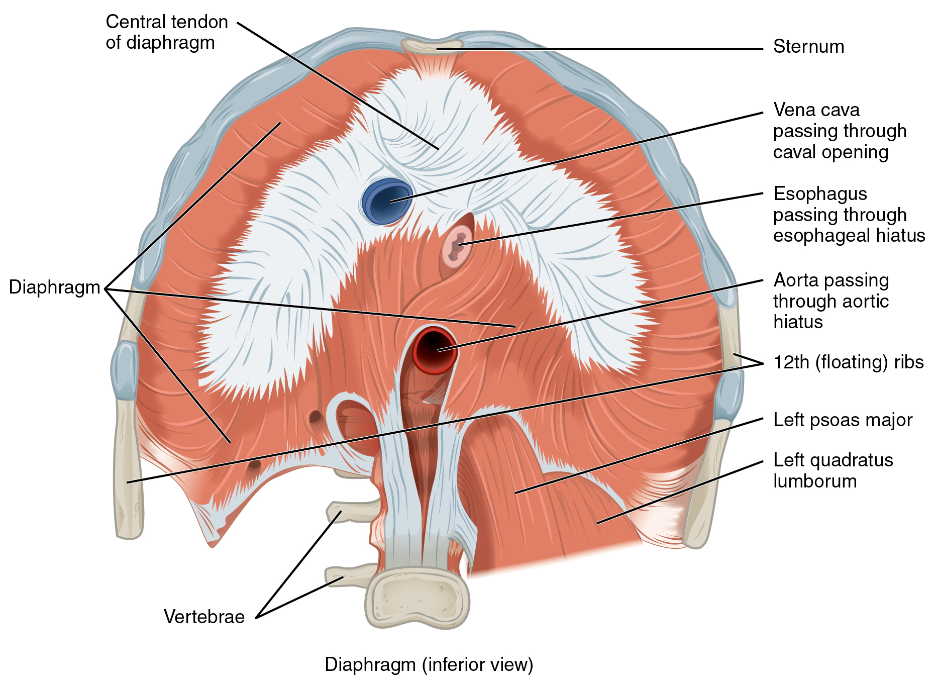
Axial Muscles of the Abdominal Wall, and Thorax · Anatomy and Physiology

Human diaphragm anatomy. Drawing of a normal human diaphragm in
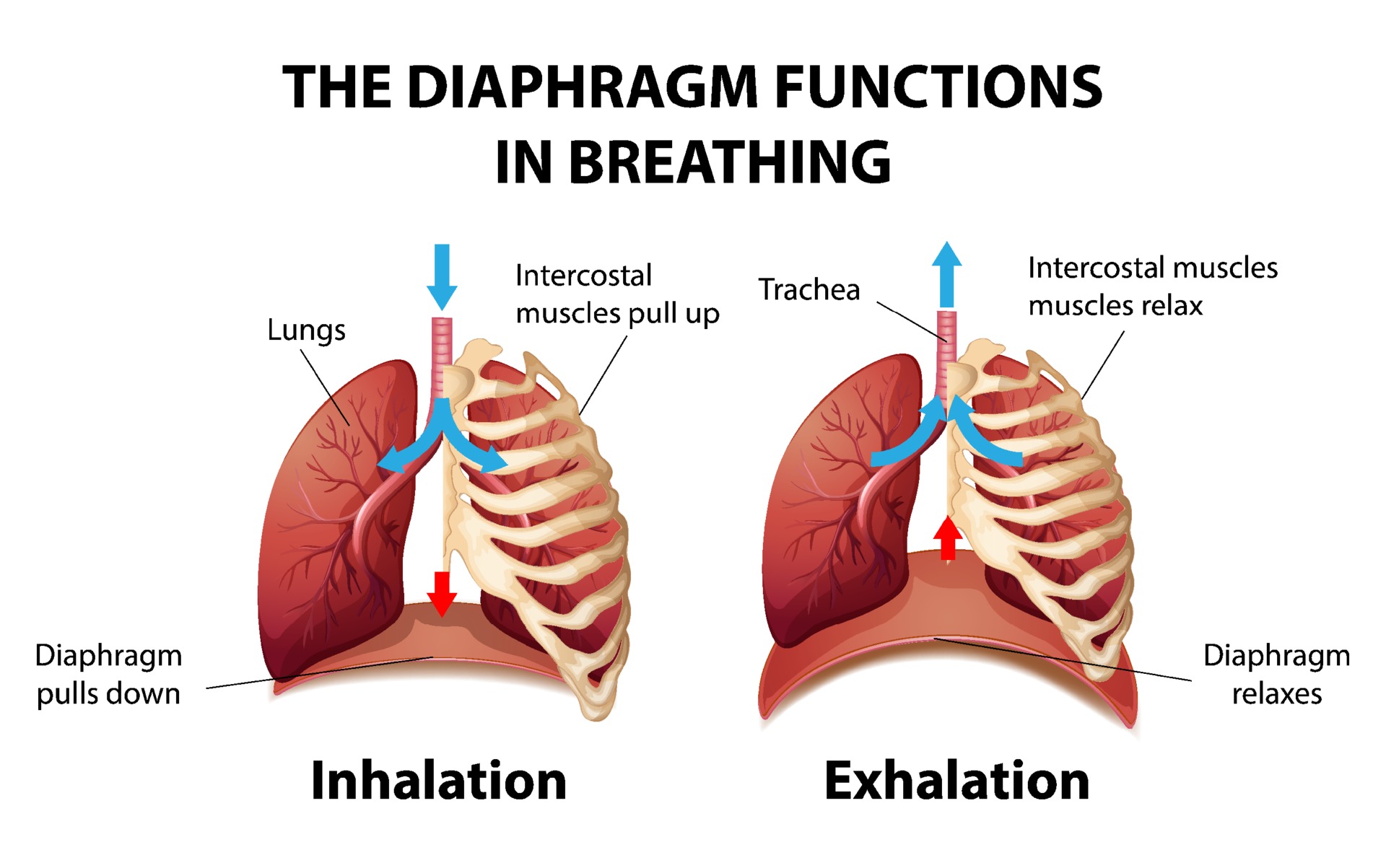
The diaphragm functions in breathing 3093606 Vector Art at Vecteezy

Diaphragm, illustration Stock Image C039/1829 Science Photo Library
Web The Diaphragm Is A Muscular Partition That Separates The Thoracic Cavity From The Abdominal Cavity In The Human Body.
This Creates A Vacuum Effect That.
Web The Diaphragm Is A Muscle That Helps You Breathe.
It Is Attached Anteriorly To The Xiphoid Process And Costal Margin, Laterally To The 11Th And 12Th Ribs, And Posteriorly To The Lumbar Vertebrae.the Posterior Attachment To The Vertebrae Is By Tendinous Bands Called The.
Related Post: