Diagram Respiratory System Drawing
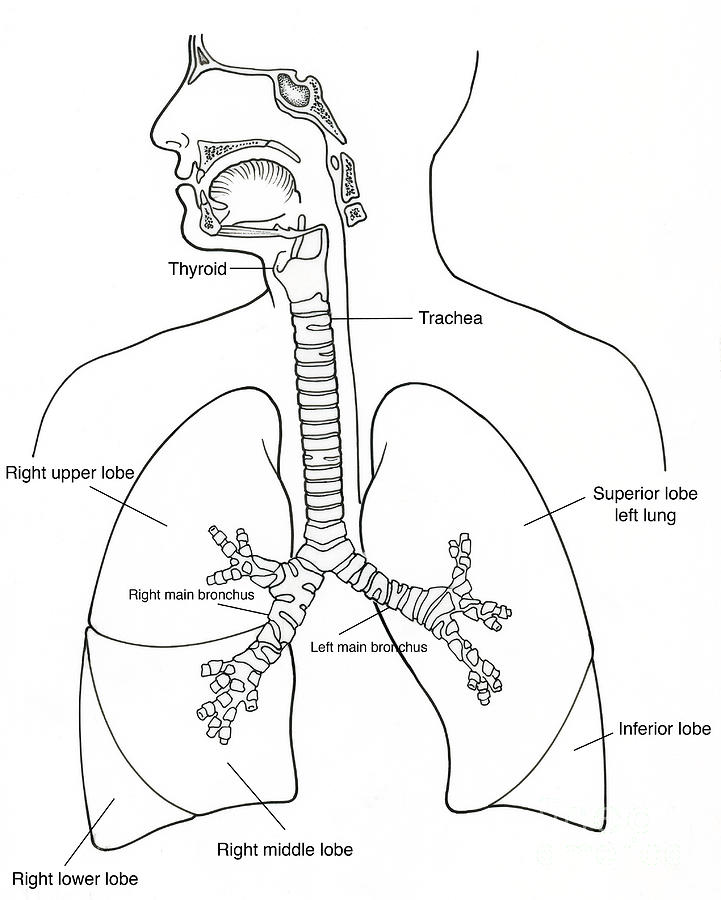
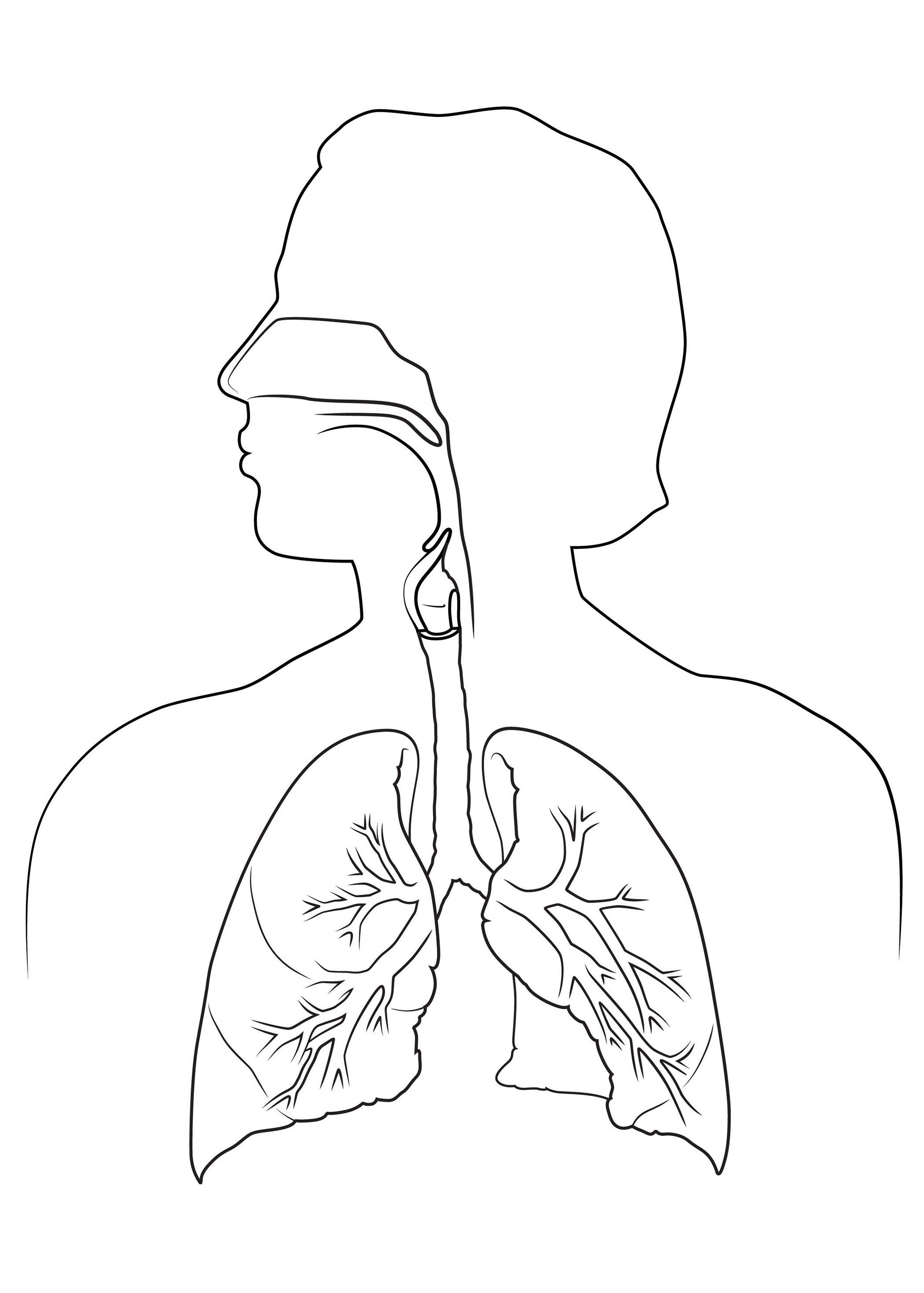
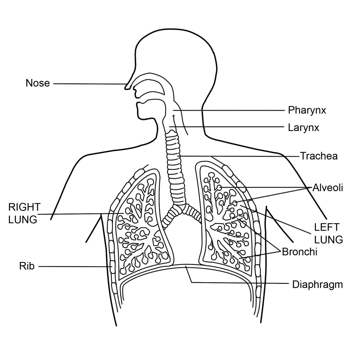
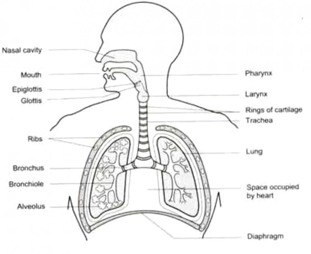
Diagram Respiratory System Drawing - Outline the thoracic cavity, neck, and head. Web learn how to draw the respiratory system of human body with this easy and fun video tutorial. The respiratory tract has two major divisions: The lower respiratory tract is. The major respiratory structures span the nasal cavity to the diaphragm. Draw the frame for the diagram. Your respiratory system is made up of your lungs, airways (trachea, bronchi and bronchioles), diaphragm, voice box, throat, nose and mouth. Functionally, the respiratory system can be divided into a conducting zone and a respiratory zone. Web hi friends,breathing is the process that brings oxygen in the air into your lungs and moves oxygen and through your body. You will also find bowman's (olfactory) glands that secrete mucus to help lubricate the mucosal layer and to dissolve odorants. Web the respiratory tract conveys air from the mouth and nose to the lungs, where oxygen and carbon dioxide are exchanged between the alveoli and the capillaries. The diagram of respiratory system. You will want this to be large enough for you to draw more detailed structures inside. Web learn how to draw diagram of human respiratory system, using a. Throat (pharynx) voice box (larynx) windpipe (trachea) large airways (bronchi) lungs. Your respiratory system is made up of your lungs, airways (trachea, bronchi and bronchioles), diaphragm, voice box, throat, nose and mouth. The diagram of respiratory system. This system also removes waste gases. The upper respiratory tract and the lower respiratory tract. 1 is lined with ciliated pseudostratified epithelial with goblet cells and this makes up the mucosal layer. Draw the frame for the diagram. Web pneumonia symptoms must include acute onset of lower respiratory illness with fever and/or cough. Web the respiratory system is made up of the organs included in the exchange of oxygen and carbon dioxide. Below, you’ll find. During internal respiration, oxygen and carbon dioxide. Choose a reference photo online or from your textbook to follow along with. Web learn how to draw diagram of human respiratory system, using a very easy to understand method. Outline the thoracic cavity, neck, and head. Below, you’ll find the respiratory system labeled and unlabeled on two. External respiration, also known as breathing, involves both bringing air into the lungs (inhalation) and releasing air to the atmosphere (exhalation). Throat (pharynx) voice box (larynx) windpipe (trachea) large airways (bronchi) lungs. Web the respiratory tract conveys air from the mouth and nose to the lungs, where oxygen and carbon dioxide are exchanged between the alveoli and the capillaries. Our. Headache, chills, shortness of breath, gi symptoms, confusion treated with antibiotics pontiac fever milder illness at least one symptom including fever, chills, myalgia, malaise, headache, fatigue, nausea/vomiting often self. 1 is lined with ciliated pseudostratified epithelial with goblet cells and this makes up the mucosal layer. Web pneumonia symptoms must include acute onset of lower respiratory illness with fever and/or. Headache, chills, shortness of breath, gi symptoms, confusion treated with antibiotics pontiac fever milder illness at least one symptom including fever, chills, myalgia, malaise, headache, fatigue, nausea/vomiting often self. The conducting zone of the respiratory system includes the organs and structures not directly involved in gas exchange. Web the nasal epithelium (figure 20.6.1 20.6. This process involves inhaling air and. Outline the thoracic cavity, neck, and head. The respiratory system is the chain of organs and tissues that helps living beings breathe properly. The respiratory system diagram shows the nose, mouth, pharynx, larynx, trachea, bronchi, and lungs. This system also removes waste gases. The organs in each division are shown in figure 16.2.2 16.2. The diagram of respiratory system. 1 is lined with ciliated pseudostratified epithelial with goblet cells and this makes up the mucosal layer. Draw the frame for the diagram. Web the respiratory system aids the body in the exchange of gases between the air and blood, and between the blood and the body’s billions of cells. The major respiratory structures span. The respiratory system functions to move air in and out of the lungs so that we can get oxygen and release carbon dioxide. The respiratory system allows air to reach the lungs, from which oxygen enters the blood and circulates to all body cells. The major respiratory structures span the nasal cavity to the diaphragm. The muscles responsible for the. The gas exchange occurs in the. 1 is lined with ciliated pseudostratified epithelial with goblet cells and this makes up the mucosal layer. Deep to this layer will be numerous bipolar cell nuclei. The respiratory system diagram shows the nose, mouth, pharynx, larynx, trachea, bronchi, and lungs. This is a great way to. Web what is respiration:the process ofreleasing energy from foodis known as respiration.it is achieved byoxidising simple food moleculeslike glucose.this processrequires oxygen.the exchange of gases in our body is brought about by the process ofbreathing.respiration takes place in themitochondriaof cell. Choose a reference photo online or from your textbook to follow along with. Web the respiratory tract conveys air from the mouth and nose to the lungs, where oxygen and carbon dioxide are exchanged between the alveoli and the capillaries. It includes the nasal passage, mouth, lungs, and blood vessels. Our lungs remove the oxygen the res. The respiratory tract in humans is made up of the following parts: Web the organs of the respiratory system form a continuous system of passages called the respiratory tract, through which air flows into and out of the body. Web the lungs are the main part of your respiratory system. Web the respiratory system aids the body in the exchange of gases between the air and blood, and between the blood and the body’s billions of cells. Your respiratory system is made up of your lungs, airways (trachea, bronchi and bronchioles), diaphragm, voice box, throat, nose and mouth. External respiration and internal respiration.
Easy Respiratory System diagram drawing How to drawing respiratory
Respiratory System Diagram Sketch Coloring Page
Respiratory System Drawing at GetDrawings Free download

Human Respiratory System 7 Download Scientific Diagram

Schematic representation of the respiratory system. Download
What is the Respiratory System Diagram and Function HubPages
Labeled Diagram Of The Respiratory System Of A Human
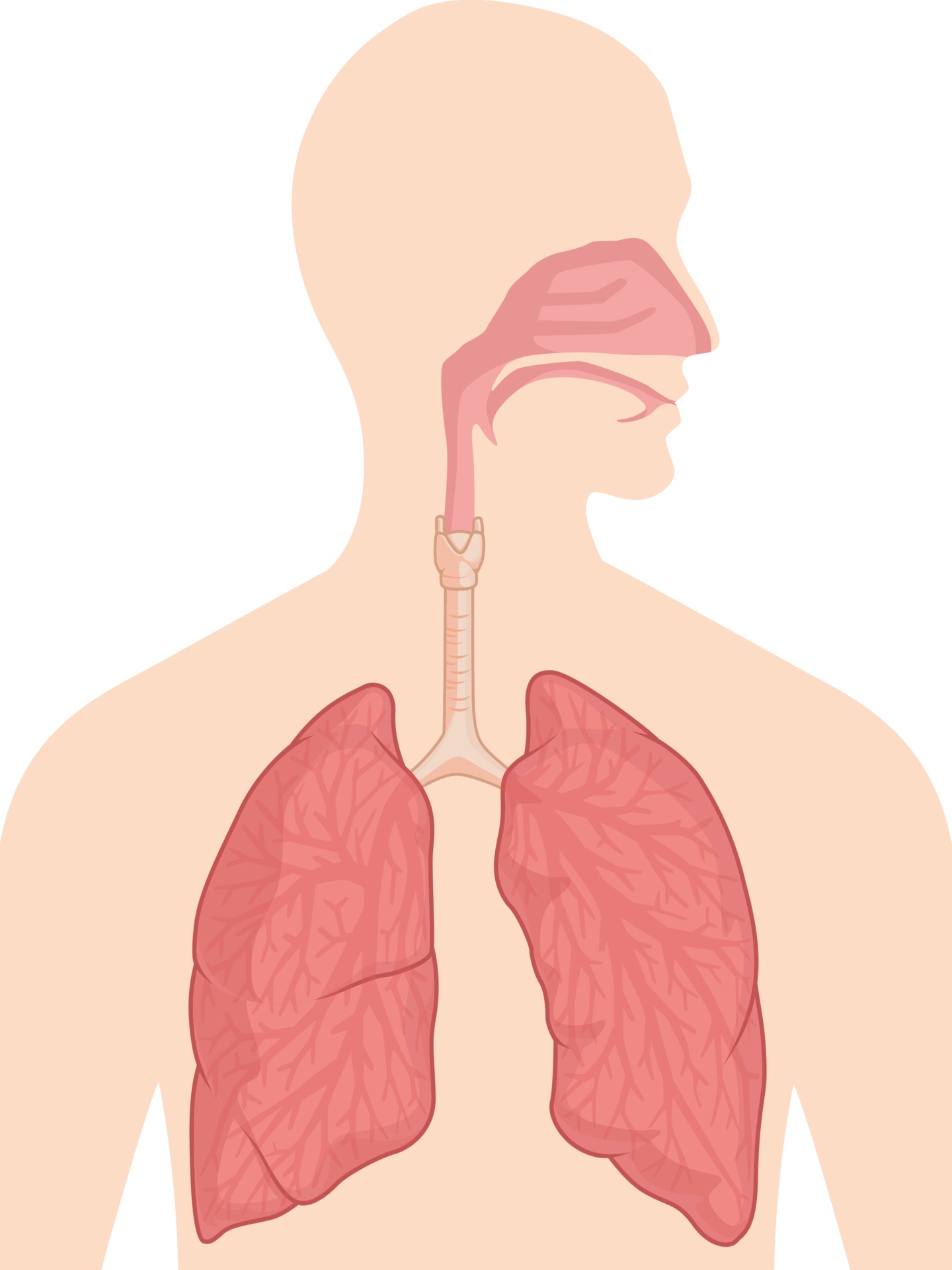
Respiratory Breathing System Body Organ Anatomy Diagram Vector Drawing

draw a labelled diagram showing the human respiratory system Brainly.in

How to draw a Human Respiratory system Diagram Drawing easy science
The Respiratory Tract Has Two Major Divisions:
Functionally, The Respiratory System Can Be Divided Into A Conducting Zone And A Respiratory Zone.
The Respiratory System Functions To Move Air In And Out Of The Lungs So That We Can Get Oxygen And Release Carbon Dioxide.
Below, You’ll Find The Respiratory System Labeled And Unlabeled On Two.
Related Post: