Chart Of Heart
Chart Of Heart - Base (posterior), diaphragmatic (inferior), sternocostal (anterior), and left and right pulmonary surfaces. In the simplest terms, the heart is a pump made up of muscle tissue. The average human heart weighs between. The movement of electrical signals across the heart is what is traced on an electrocardiogram (ekg). Important questions about the human heart. The surfaces and borders of the heart. Web home health conditions and diseases. It serves as an indication of your general fitness. The right atrium, left atrium, right ventricle, and left ventricle. The heart is a conical hollow muscular organ situated in the middle mediastinum and is enclosed within the pericardium. A typical heart is approximately the size of your fist: On may 29, south africans head to the polls. Web the aha heart disease and stroke statistical update presents the latest data on a range of major clinical heart and circulatory disease conditions (including stroke, brain health, complications of pregnancy, kidney disease, congenital heart disease, rhythm disorders, sudden cardiac arrest,. The atria are smaller than the ventricles and have thinner, less muscular walls than the ventricles. Web anatomy of the heart. Web south africa elections 2024 explained in maps and charts. The movement of electrical signals across the heart is what is traced on an electrocardiogram (ekg). Web home health conditions and diseases. You have two chambers on the top (atrium, plural atria) and two on the bottom (ventricles), one on each side of your heart. The right atrium, left atrium, right ventricle, and left ventricle. 12 cm (5 in) in length, 8 cm (3.5 in) wide, and 6 cm (2.5 in) in thickness. On may 29, south africans head to the polls.. Learn more about the heart in this article. The valves of the heart. Welcome to the anatomy of the heart made easy! You have two chambers on the top (atrium, plural atria) and two on the bottom (ventricles), one on each side of your heart. A typical heart is approximately the size of your fist: It is divided by a partition (or septum) into two halves. The atria act as receiving chambers for blood, so they are connected to the veins that carry blood to the heart. In addition to reviewing the human heart anatomy, we will also discuss the function and order in which blood flows through the heart. Base (posterior), diaphragmatic (inferior), sternocostal. It is divided into the left and right sides by a muscular wall called the septum. Blood is transported through the body via a complex network of veins and arteries. Important questions about the human heart. 12 cm (5 in) in length, 8 cm (3.5 in) wide, and 6 cm (2.5 in) in thickness. You have two chambers on the. Web the normal resting heart rate (when not exercising) for people age 15 and up is 60 to 100 beats per minute (bpm). It is divided by a partition (or septum) into two halves. The right atrium, left atrium, right ventricle, and left ventricle. Practise labelling the human heart diagram. The heartbeat drives the transport of blood throughout the body,. It also has several margins: On may 29, south africans head to the polls. The halves are, in turn, divided into four chambers. Practise labelling the human heart diagram. After 30 years of dominance, the anc faces its toughest election yet, needing 50 percent to. Includes the anatomy of the heart and an animation quiz at the end in order to test your knowledge! The heart is made of three layers of tissue. Base (posterior), diaphragmatic (inferior), sternocostal (anterior), and left and right pulmonary surfaces. It also has several margins: The halves are, in turn, divided into four chambers. Web the cardiovascular system. It is covered by a sack termed the pericardium or pericardial sack. Two atria and two ventricles. Although the heart is a. It controls the electrical impulses that cause your heart to beat and their conduction, which organizes the beating of your heart. What should your heart rate be when working out, and how can you keep track of it? The surfaces and borders of the heart. Like all muscle, the heart needs a source of energy and oxygen to function. New 3d rotate and zoom. The valves of the heart. The electrical system of the heart is critical to how it functions. Updated on april 05, 2020. Blood is transported through the body via a complex network of veins and arteries. It also has several margins: Web your heart has four separate chambers. Web the heart has five surfaces: The superior vena cava carries blood from your upper body. Base (posterior), diaphragmatic (inferior), sternocostal (anterior), and left and right pulmonary surfaces. Save time with a video! Web the normal resting heart rate (when not exercising) for people age 15 and up is 60 to 100 beats per minute (bpm). Learn all about the heart, blood vessels, and composition of blood itself with our 3d models and explanations of cardiovascular system anatomy and physiology.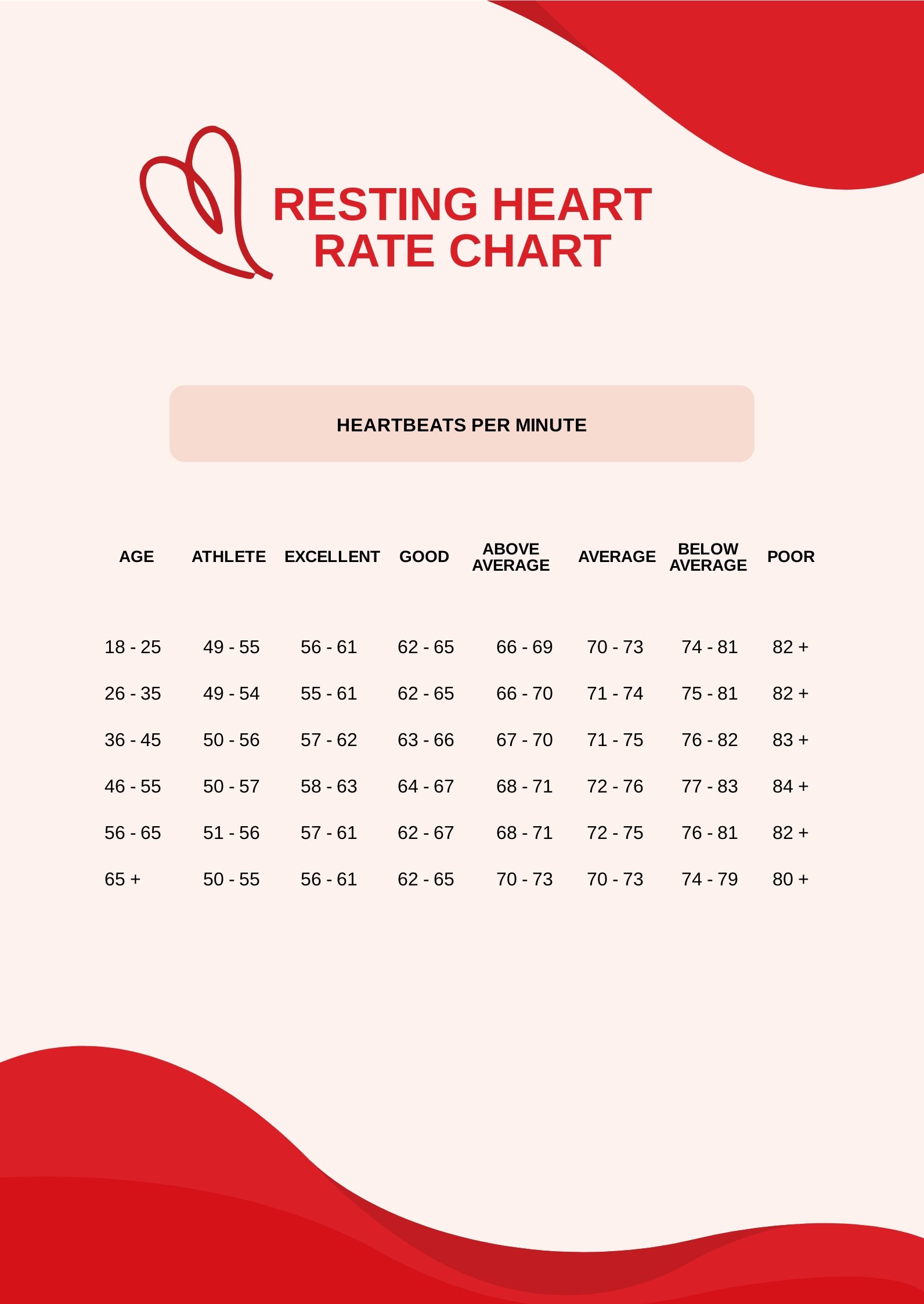
Heart Rate Recovery Chart By Age
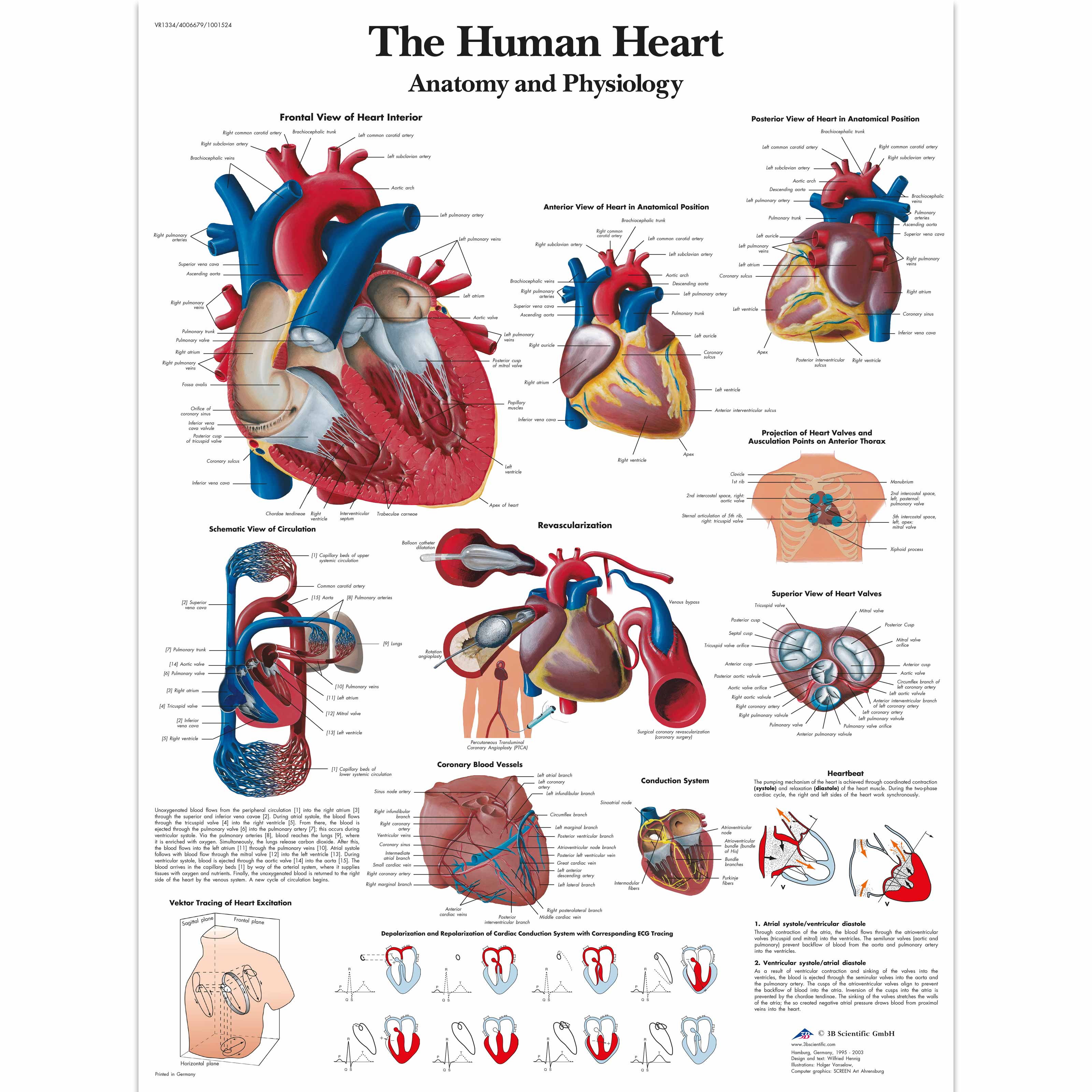
The Human Heart Chart Anatomy and Physiology 4006679 VR1334UU

Blood Pressure and Heart Rate Chart Free Download
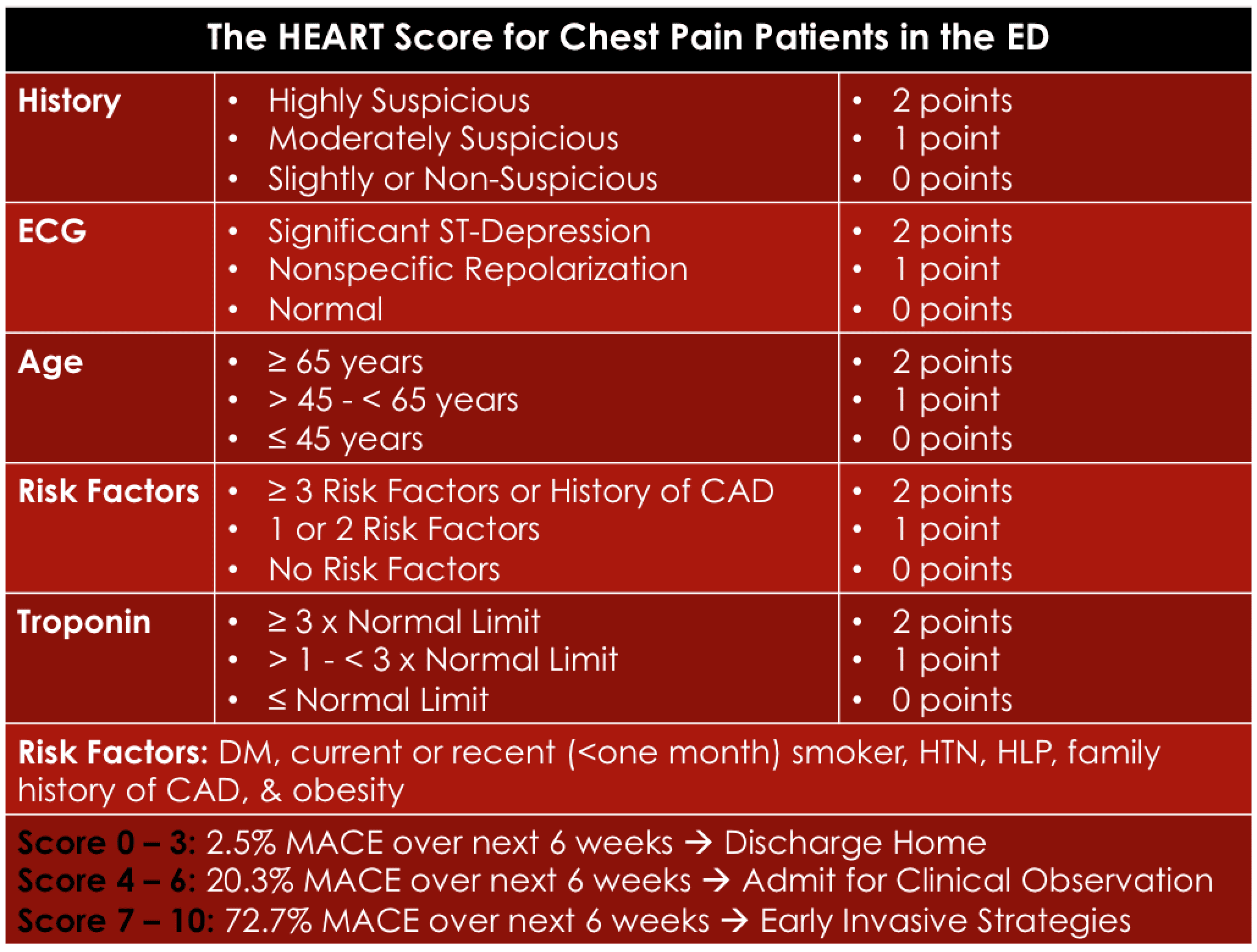
The HEART Score REBEL EM Emergency Medicine Blog
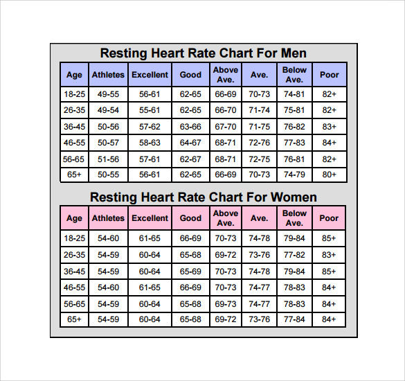
FREE 12+ Sample Heart Rate Chart Templates in PDF MS Word Excel
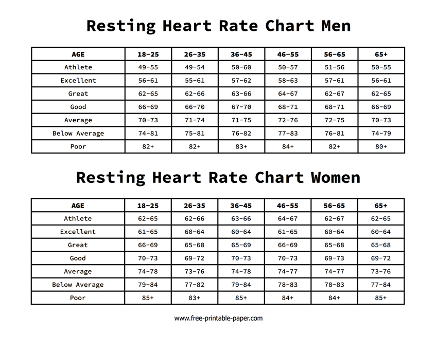
Resting Heart Rate Chart
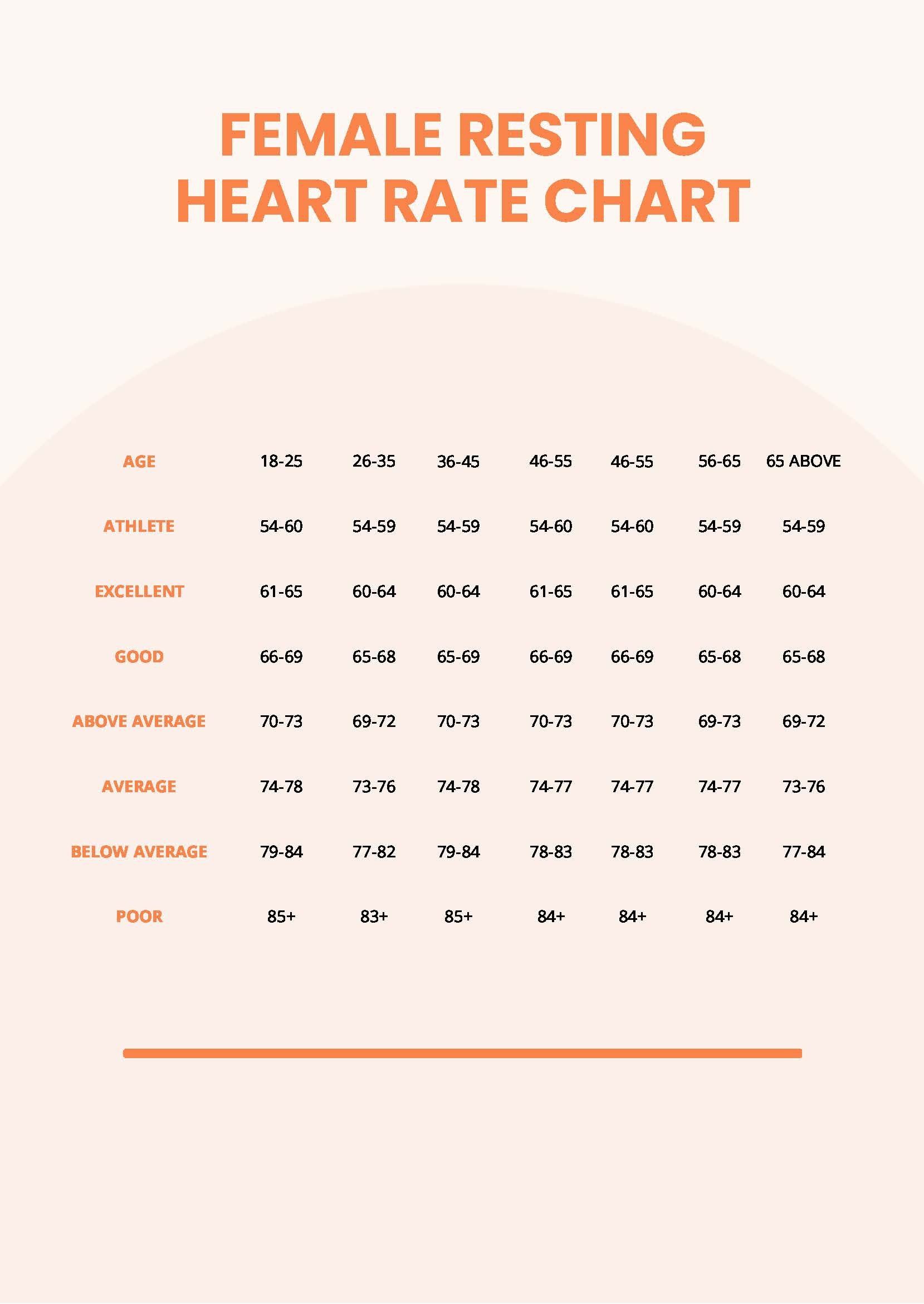
Heart Rate Chart By Age And Gender PDF
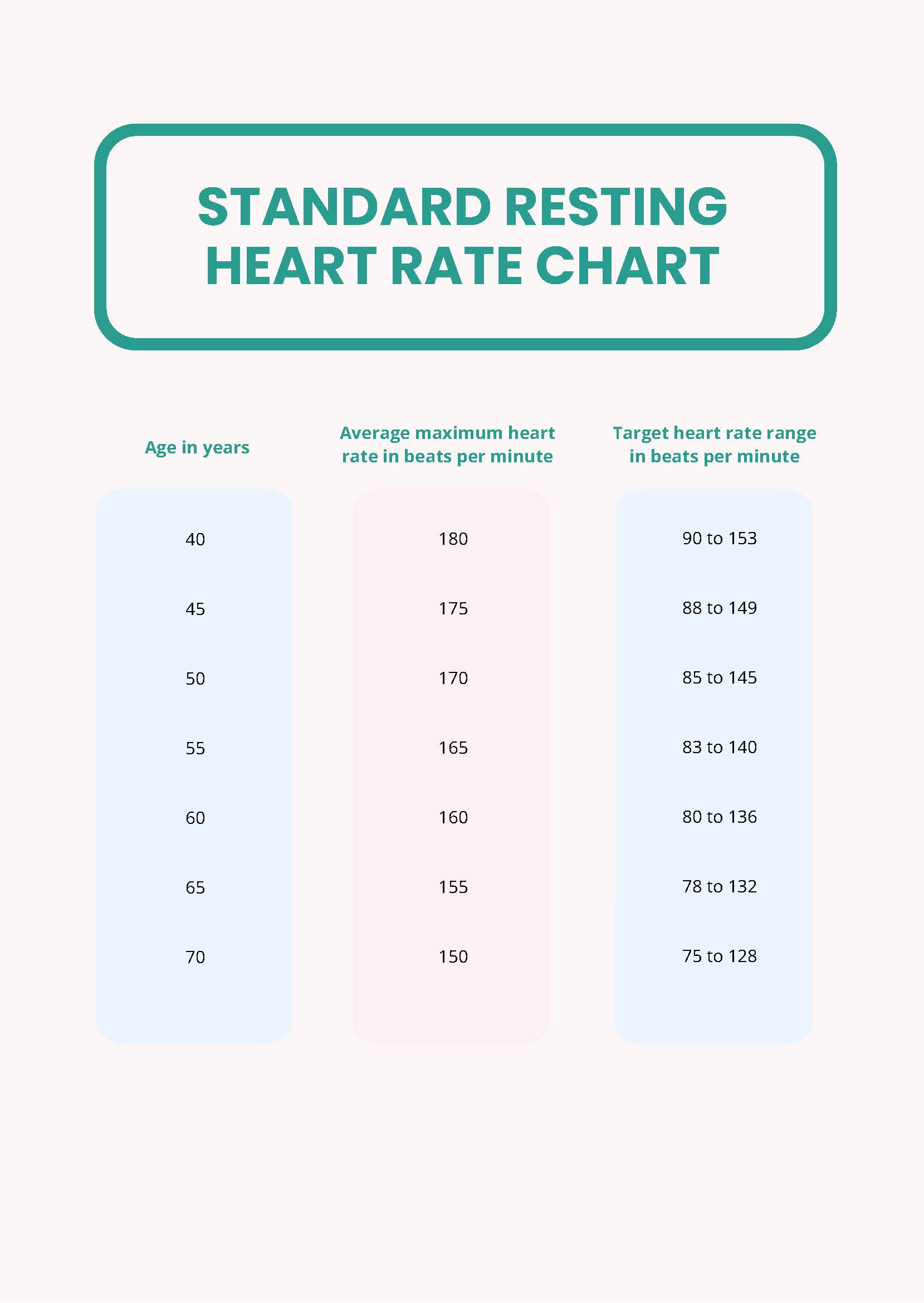
Low Resting Heart Rate Chart in PDF Download
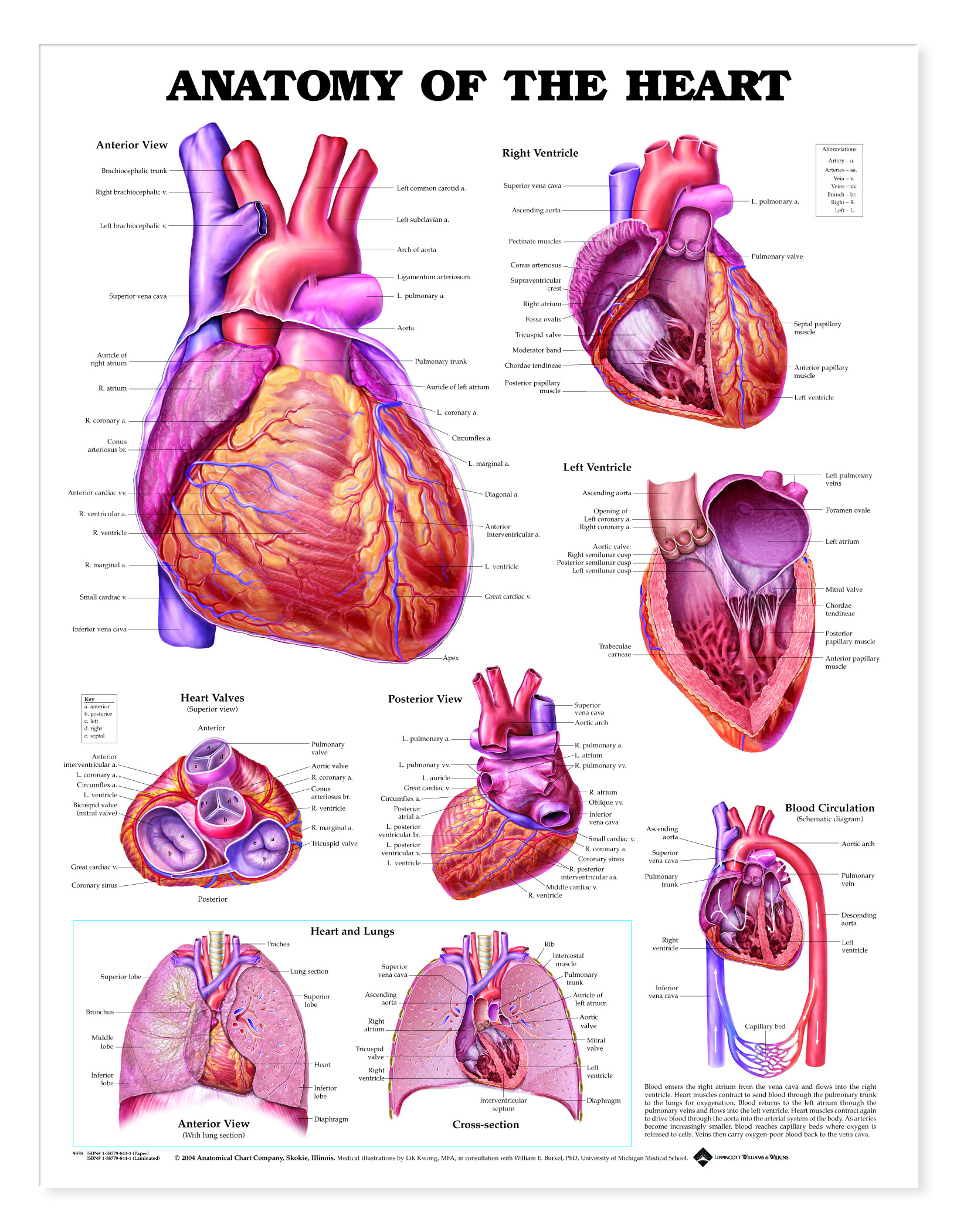
Anatomy of the Heart Charts 1944
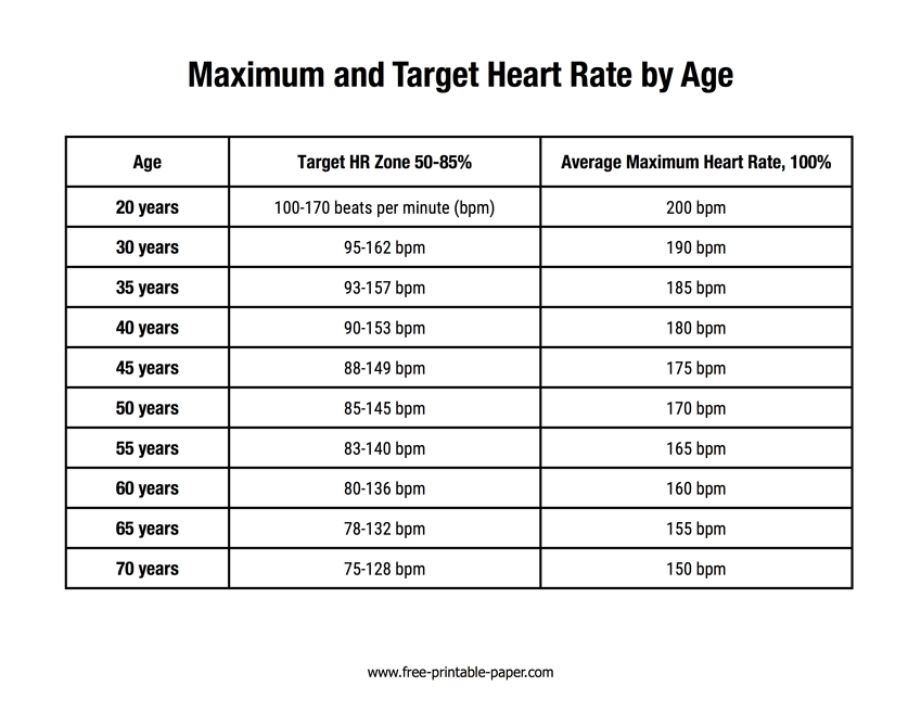
Heart Rate Chart Free Printable Paper
Web The Cardiovascular System.
Right, Left, Superior, And Inferior:
The Heartbeat Drives The Transport Of Blood Throughout The Body, Which Provides Oxygen And Nutrients To All The Body’s Cells, Tissues, And Organs.
It Controls The Electrical Impulses That Cause Your Heart To Beat And Their Conduction, Which Organizes The Beating Of Your Heart.
Related Post: